Download
1 / 42
680 likes | 1.73k Views
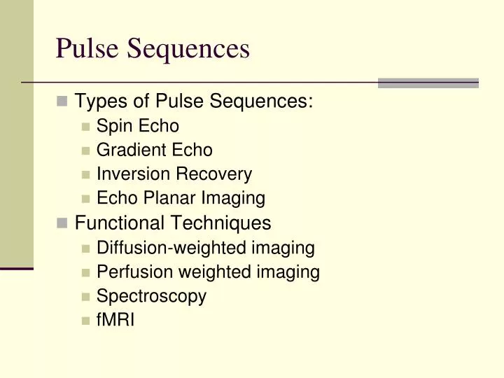
Pulse Sequences. Types of Pulse Sequences: Spin Echo Gradient Echo Inversion Recovery Echo Planar Imaging Functional Techniques Diffusion-weighted imaging Perfusion weighted imaging Spectroscopy fMRI. Pulse Sequences. Spin Echo (SE) (Conventional Spin Echo)

E N D
Pulse Sequences • Types of Pulse Sequences: • Spin Echo • Gradient Echo • Inversion Recovery • Echo Planar Imaging • Functional Techniques • Diffusion-weighted imaging • Perfusion weighted imaging • Spectroscopy • fMRI
Pulse Sequences Spin Echo (SE) (Conventional Spin Echo) RF pulse sequence with a 90o excitation pulse followed by a 180o rephasing or refocusing pulse to eliminate field inhomogeneity and chemical shift effects. • Main Points: 1) 90o excitation pulse 2)180o rephasing pulse
Pulse Sequences Spin Echo (SE)
Pulse Sequences Spin Echo (SE)
Pulse Sequences Spin Echo – Multiple Echo
Pulse Sequences Spin Echo – Multi-echo
Pulse Sequences Spin Echo Pulse Sequence • T1 weighted images • Short TR (300 ms – 700 ms) • Short TE (10 ms – 30 ms) • T2 weighted Images • Long TE ( > 80 ms) • Long TR ( > 2000 ms)
Pulse Sequences T1WI vs. T2WI ____________________________________ *Short TR *Long TE Short TE Long TR T1WI T2WI
Pulse Sequences Proton (Spin) Density Weighted • Density of Resonating spins in a given volume (e.g., tissue). • The higher the concentration of spin density, the higher (brighter) the received signal. • Long TR (1000 ms – 2000 ms) • Short TE (10 ms – 30 ms)
Pulse Sequences Spin Echo • T1 – weighted image (short TR, short TE) • Proton Density image (long TR, short TE) • T2 – weighted image (long TE, long TR)
Pulse Sequences Rapid Acquisition with Relaxation Enhancement • Also known as RARE • Fast Spin Echo (FSE) – GE, Picker, Toshiba & Hitachi • Turbo Spin Echo (TSE) – Siemens & Philips • One advantage is speed without loss of S/N. In CSE, if acquisition time is reduced by 50%, the S/N is reduced by 40%
Pulse Sequences • Fast Spin Echo
Pulse Sequences RARE
Pulse Sequences Fast (Turbo) Spin Echo • Echo Train Length (ETL) or Turbo Factor (TF) • Effective Echo Time (ETE) • Echo Train Spacing (ETS)
Pulse Sequences Rapid Acquisition with Relaxation Enhancement • Acquires multiple echoes per excitation at different phase encoding steps – echo train length (ETL) • The number of echos acquired per excitation pulse is referred to as echo train length • TE is referred to as “Effective TE” (ETE)
Pulse Sequences Rapid Acquisition with Relaxation Enhancement • Time between each echo (phase encoding step) is referred to as echo train spacing (ETS) • TE is primarily responsible for image contrast. • RARE reduces the amount of excitation pulses. • Scan Time – (TR x Gpe x NEX) / ETL
Pulse Sequences Fast Spin EchoScan Time = (TR x Gpe X NEX) / ETL TR = 2000 ms (2000 x 256 x 4) / 8 Gpe = 256 2000 x 256 x 4 = 2048000 NEX = 4 2048000 / 1000 = 2048 seconds ETL = 8 2048 / 60 (convert seconds to minutes) = 34.13 minutes 34.13 minutes / 8ETL = 4.26 minutes
Pulse Sequences Fast Spin Echo • Advantage • Reduced Scan Time • Disadvantage • Possible flow and motion artifacts • Fat may be brighter on FSE than CSE • Image blur – Increased TF or ETL
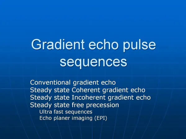
Pulse Sequences • Gradient Echo (GE) Generally uses an excitation flip angle (FA) of less than 90o degree and a gradient reversal to rephase the protons • Main Points: 1) Variable Flip Angle (FA) 2) Gradient reversal
Pulse Sequences • Gradient Echo
Pulse Sequences Advantages: Gradient Echo • Much shorter scan times than SE pulse sequences • Low FA allows for faster recovery of longitudinal magnetization • Gradients rephase faster than 180o RF pulses • TR and TE values are shorter than spin echo pulse sequences
Pulse Sequences Disadvantages: Gradient Echo • Susceptible to magnetic field inhomogeneities • Contain magnetic susceptibility artifacts • T2* weighting
Pulse Sequences Fast Spin Echo (Advantage over GRE) • FSE uses a 180 degree pulse to eliminate susceptibility artifacts. • Heavy T2 – weighted images cannot be easily acquired with GRE
Pulse Sequences • Gradient Echo: Weighting • T1 Weighting - 1o controlled by FA and TR • T2* Weight – 1o controlled by TE
Pulse Sequences • Gradient Echo Pulse Sequence • T1 – Weighted • Large Flip Angle • Short TR • Short TE • T2* - weighted • Small Flip Angle • Long TR • Long TE • Proton Density Weighted • Small Flip Angle • Long TR • Short TE
Pulse Sequences Inversion Recovery Sequence consisting of an initial 180o RF pulse to invert the magnetization, followed by a spin-echo (90o to 180o) or gradient sequence.
Pulse Sequences Inversion Recovery
Pulse Sequences Inversion Recovery • FLAIR – Nulls CSF • STIR – Nulls FAT STIR FLAIR
Pulse Sequences • Dixon
Pulse Sequences Null Point
Pulse Sequences Echo Planar Imaging (EPI) • Allows data to be collected and reconstructed in less than a second • Stronger and faster gradients (slew rate) required • Data collected along GFE (readout) direction
Pulse Sequences • Echo Planar Imaging
Functional Techniques • Diffusion-weighted image (DWI) • Technique used to measure the motion of water molecules • Areas with increased diffusion within a tissue show greater signal loss • Terms such as – b-value, Apparent diffusion coefficient (ADC) • Clinical applications – acute cerebral infarcts (ischemic areas) appear bright
Functional Techniques • Perfusion-weighted image (PWI) • Technique used to measure the flow of blood though the capillary region of an organ or tissue • Dynamic Susceptibility Contrast (DSC) uses gadolinum as a tracer • Arterial Spin Labeling (ASL) noninvasive • Terms - Cerebral Blood Flow (CBF), Mean transit Time (MTT) and Cerebral Blood Volume (CBV)
Functional Techniques • Spectroscopy (MRS) • Technique that provides chemical information about a tissue • Nuclei such as 1H, 31P, 13C, 23Na, 39K, 19F • Methods used: • Stimulated echo acquisition mode (STEAM) • Point-resolved spectroscopy (PRESS) • Image-selected in vivo Spectroscopy (ISIS) • Chemical shift imaging (CSI) a.k.a. magnetic resonance spectroscopic imaging (MRSI)
Functional Techniques • Functional MRI (fMRI) • Term used to describe any technique that evaluates brain physiology rather than anatomy • fMRI specifically used to map brain activity • Areas of interest – motor cortex, visual cortex • Blood oxygenation level-dependent (BOLD)
Functional Magnetic Resonance Imaging fMRI BOLD: Blood Oxygen Level Dependence fMRI is the most common for research purposes Measures the hemodynamic response (amount of blood flow) related to neural activity in the brain Increase in magnetic susceptibility when blood is oxygenated Direct correlation between brain activity and cerebral blood flow has been observed, but unsure of exact underlying mechanisms Usually uses a T2* weighted contrast TR: 2-4s 2-4mm spatial resolution (better with 3-9T)
Methods Two common types of experimental design Resting state: Participant has no task, just lies still in the scanner Experimental: Participants are presented with tasks or stimuli at approximately 5s intervals Or block design due to poor temporal resolution 5s + 5s … to 2 min Repeat w Next Block of Images
Methods- cont’d Although commonly thought of as a direct measure of brain activity, fMRI usually identifies relative differences in brain activity Correlation does NOT equal causality Subtraction Technique EX: Response to images of smoking Smokers Nonsmokers Difference - =
Clinical Utility fMRI is not currently used for diagnostic purposes Used to identify functional maps of neural networks & research relative deficiencies in brain function New software allows for the quantification of network activity via Independent Components Analysis Differences in network connectivity has been associated with certain disorders and fMRI should be capable of accurate diagnosis within the next decade Allows early identification and treatment of disordered individuals (e.g. Schizophrenia and Alzheimers) Visual Network Default Mode Network
Other Functional Techniques Diffusion based fMRI Contrast MR Arterial Spin Labeling Magnetic Resonance Spectroscopic Imaging Electroencephalography Although it has great spatial resolution, fMRI has poor temporal resolution of neuronal activity Combined EEG-fMRI may allow quantification of activity at sub-ms and sub-mm resolution