Download
1 / 140
1.69k likes | 2.24k Views
Digestive System. http://www.madras.com. Much of the text material is from, “Principles of Anatomy and Physiology, 12th edition” by Gerald J. Tortora and Bryan Derrickson (2009). I don’t claim authorship. Other sources are noted when they are used.

E N D
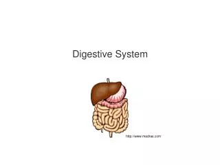
Digestive System http://www.madras.com
Much of the text material is from, “Principles of Anatomy and Physiology, 12th edition” by Gerald J. Tortora and Bryan Derrickson (2009). I don’t claim authorship. Other sources are noted when they are used. Mapping of the lecture slides to the 13th edition is provided in the supplement.
Outline • Introduction • Overview • Tissue layers • Nervous system control • Upper gastrointestinal tract • Lower gastrointestinal tract • Phases of digestion • Aging
Food and Digestion • Food supplies nutrients for metabolism, tissue growth, and repair of damaged tissues. • Food is the only source of chemical energy that can be used by the body. • Most of the food we eat consists of molecules—such as proteins, lipids, and complex sugars—that are too large to be used by cells. • Food is broken-down into smaller molecules by digestive processes. Chapter 24, page 921
Food and Digestion (continued) • The organs involved in the digestion and absorption of food are known as the digestive system. • The digestive system is a tubular structure that extends from the mouth to the anus. • Its close working relationship with the cardiovascular system to assure the availability of chemical energy to meet the body’s metabolic needs. Chapter 24, page 921
Dietary Guidelines http://www.hirzel.com See the U.S. Department of Agriculture website, http://www.mypyramid.gov.
Definitions • The medical specialty for the diagnosis and treatment of diseases of the esophagus, stomach, and intestines is known as gastroen-terology. • The medical specialty for the rectum and anus is called proctology. Chapter 24, page 921
Gastrointestinal Tract • The digestive system consists of the gastrointestinal (GI) tract and its accessory digestive organs. • The GI tract is also known as the alimentary canal, which is a term we won’t use in this lecture. • It includes the mouth, most of the pharynx (throat), esophagus, stom-ach, small intestine, and large intestine. • The end to end length of the GI tract is about 5 to 7 meters in a living person. Figure 24.1 Chapter 24, page 922
Accessory Digestive Organs • The accessory organs include teeth, tongue, salivary glands, liver, gall bladder, and pancreas. • Teeth support the mechanical breakdown of food by chewing, while the tongue assists in chewing and swallowing. • All other accessory organs do not come into direct contact with food. • They produce or store secretions to further the chemical breakdown of food, as will be discussed. Figure 24.1 Chapter 24, page 922
Digestive System http://www.pediatricfeeding.org
Roles of Muscles • Smooth muscle contractions in the wall of the GI tract mechanically breakdown food by churning and propelling it along its hollow struc-ture. • Muscle contractions also help dissolve foods by mixing them with fluids secreted into the GI tract that chemically breakdown the food molecules. • The contractions involve segmentations and peristalsis, to be dis-cussed later in this lecture. Chapter 24, page 922
Basic Processes • The six basic processes of the digestive system are: • Ingestion • Secretion • Mixing and propulsion • Mechanical and chemical digestion • Absorption • Defecation Chapter 24, page 923
Basic Processes (continued) • Ingestion involves eating and drinking to take foods and liquids into the mouth. • Secretion involves the release of water, acids, buffers, and enzymes into the lumen of the GI tract. • Mixing and propulsion of food through the GI tract is accomplished by the alternating contraction and relaxation of smooth muscle in the walls of the GI tract. Chapter 24, page 923
Basic Processes (continued) • Mechanical and chemical digestion break food into smaller molecules to aid in their absorption. • Absorption involves the passage of small molecules into the epithelial cells lining portions of the GI tract. • The molecules pass into blood or lymph, and circulate to the liver and cells throughout the body. • A few substances can be absorbed without chemical digestion— they include vitamins, ions, cholesterol, and water. Chapter 24, page 923
Basic Processes (continued) • Defecation is the elimination of materials from the digestive tract through the anus. • The materials include waste products, indigestible substances, undigested food, bacteria, and cells from the inner lining of the GI tract. • The eliminated materials are known as feces. Chapter 24, page 923
Tissues • The wall of the GI tract—from the esophagus to the anal canal—has four layers of tissues. • From the lumen to the outer surface of the GI tract they consist of the mucosa, submucosa, muscularis, and serosa. Figure 24.2 Chapter 24, page 924
Tissues (continued) http://www.med.howard.edu
Skeletal Muscle • The muscularis of the mouth, pharynx, and upper parts of the eso-phagus consists of skeletal muscle for voluntary swallowing. • Skeletal muscle also forms the external anal sphincter for voluntary control of defecation. Chapter 24, page 925
Smooth Muscle • The muscularis is made up of smooth muscle in the rest of the GI tract. • Smooth muscle is typically organized into two layers: 1) an inner layer of circular fibers and 2) an outer layer of longitudinal fibers. • Involuntary contractions of the smooth muscle mechanically break-down food, mix it with digestive secretions, and propel it along the GI tract. • Between the two muscle layers are an intricate network of neurons known as the myenteric plexus, which is part of the enteric nervous system. Chapter 24, page 925
Innervation • The GI tract is controlled by nerve fibers from the enteric nervous system (ENS) and the autonomic nervous system. • The ENS consists of about 100 million neurons extending from the eso-phagus to the anus. • While the neurons of the ENS can function independently, they are also regulated by the parasympathetic and sympathetic divisions of the ANS. Chapter 24, page 925
Enteric Nervous System • The ENS is composed of the myenteric plexus and submucosal plexus. • Plexuses are complex networks of motor neurons, interneurons, and sensory neurons. • The myenteric plexus controls motility of the GI tract through the innervation of the circular and longitudinal smooth muscles in the walls of the tact. Motility = motion or movement. Figure 24.3 Chapter 24, page 925
Enteric Nervous System (continued) http://nutrition.mattters.com
Enteric Nervous System (continued) • The submucosal plexus innervates the accessory organs to control their secretions. • Interneurons within the ENS interconnect the two plexuses to enable coordinated control of their functions. • Sensory neurons, a type of interoreceptor, are located in the mucosal epithelium of the GI tract. Figure 24.3 Chapter 24, page 925
Autonomic Nervous System • The vagus nerve (X) supplies parasympathetic fibers to much of the GI tract. • The lower half of the large intestine, however, is innervated by para-sympathetic fibers originating in the sacral section of the spinal cord. • Parasympathetic stimulation increases gastric motility and GI secre-tions by increasing the activity of neurons in the ENS. Chapter 24, page 925
Autonomic Nervous System (continued) • Sympathetic fibers innervate the ENS through the thoracic and lumbar regions of the spinal cord. • Sympathetic stimulation decreases motility and GI secretions by inhibiting the activity of neurons in the ENS. • Physical activity and emotions including anger, fear, and anxiety can slow the digestive process. Chapter 24, page 925
Mouth • The mouth is also known as the oral cavity or buccal cavity (we will use the first term). • Structural details, including of the teeth and tongue, can be found in the textbook. • The teeth aid in mechanical digestion, or mastication, and the tongue in swallowing, or deglutition. • In this section we focus primarily on the role of the salivary glands of the mouth. Figure 24.5 Chapter 24, page 928
Salivary Glands • The mucous membrane of the mouth and tongue have salivary glands that open directly or indirectly via short ducts into the oral cavity. • Salivary glands secrete a fluid called saliva. • Just enough saliva is usually secreted to keep the mucous membranes of the mouth and pharynx moist, and to cleanse the mouth and teeth. • Salivary secretions increase before or when food enters the mouth to lubricate, dissolve, and begin its chemical breakdown by the digestive system. Chapter 24, page 929
Saliva • Saliva consists of about 99.5 percent water and 0.5 percent solutes. • The solutes—in ionic form—include sodium, potassium, chloride, bi-carbonate, and phosphate. • Mucus, urea and uric acid, immunoglobulin A, bacterial enzymes, and salivary amylase are also usually found in saliva. • Not all salivary glands secrete the same composition and percentages of solutes. Salivary amylase = an enzyme that breaks-down starch molecules. Chapter 24, page 930
Saliva (continued) • The water in saliva provides a medium for dissolving foods so that: • Food can be tasted via gustatory or taste receptors in the tongue. • Chemical digestion can begin. • The solutes have specific functions including buffering of acidic foods by bicarbonate and phosphate ions, and activation of salivary amylase by chloride ions. Gustatory receptors = taste receptors in taste buds that interact with chemicals in food to produce action potentials that re transmitted to the brain. Chapter 24, page 930
Saliva (continued) • Immunoglobulin A helps keep microbes in the external environment from penetrating the mucous membrane. • Lysozyme—an enzyme—kills but does not completely eliminate bac-teria in the oral cavity. • Mucus lubricates food so that it is moved more easily in the mouth, formed into a ball, and swallowed. Chapter 24, page 930
Salivation • About 1000 to 1500 mL (1.0 to 1.5 L) of saliva is secreted each day, although the amount can vary. • Saliva moistens the oral cavity, and when swallowed, helps moisten the esophagus. • Saliva is eventually reabsorbed to minimize fluid loss from the body. • If the body is dehydrated, the secretion of saliva ceases, contributing to a conscious sensation of thirst. Chapter 24, page 931
ANS Control • Salivation—the secretion of saliva—is controlled by the autonomic nervous system. • Parasympathetic activity produces a continuous secretion of mod-erate amounts of saliva. • Sympathetic activity decreases salivary secretion to produce a dry mouth, such as in fear and anxiety. Chapter 24, page 931
Sensory and Cognitive Stimulation • The feel and taste of food stimulates salivary gland secretions by activating the gustatory receptors of the tongue. • Nerve impulses are conveyed to salivary nuclei in the brainstem. • Parasympathetic control is via cranial nerves VII and IX to stimu-late the secretion of saliva. • The smell, sight, or thought of food can also stimulate the secre-tion of saliva. Cranial nerve VII = facial. Cranial nerve IX = glossopharyngeal. Chapter 24, page 931
Pavlov’s Dogs http://nournours.files.wordpress.com Classical conditioning of the salivary response.
Digestion—Mouth • Mechanical digestion in the mouth involves chewing or mastication in which food is manipulated by the tongue, ground by teeth, and mixed with saliva. • The food is reduced to a soft and flexible mass known as a bolus. • Certain foods begins to dissolve in saliva since the salivary enzymes react with the food molecules. Table 24.1 Chapter 24, page 931
Digestion—Mouth (continued) • While most carbohydrates we eat are starches, only monosaccharides (single sugar molecules) can be absorbed into the blood. • The enzymes salivary amylase and lingual lipase contribute to chemical digestion in the mouth. • Salivary amylase starts the chemical digestion of starches into smaller molecules. • The process continues in the stomach and small intestine to form mono-saccharides. Chapter 24, page 931
Digestion—Mouth (continued) • Lingual lipase, an enzyme secreted by the lingual glands in the tongue, is activated in the acidic environment of the stomach. • The enzyme starts the process of chemical digestion of triglycerides into diglycerides and fatty acids. Triglyceride = three fatty acid chains covalently bonded to a glycerol molecule. Diglyceride = two fatty acid chains covalently bonded to a glycerol molecule. Fatty acid = an organic compound composed of a carbon chain with hydrogen atoms attached, and an acid group at each end. Chapter 24, page 931
Pharynx • When food is swallowed, it passes from the mouth into the pharynx or throat. • The pharynx is lined by mucous membrane—it also contains skele-tal muscle. • Skeletal muscle contractions of the pharynx propel the bolus of food into the esophagus. Chapter 24, page 934
Esophagus • The esophagus is a tube about 25 cm long located posterior to the trachea. • The esophagus: • Begins at the inferior end of the laryngopharynx, • Pierces the diaphragm through the esophageal hiatus, and • Ends in the superior portion of the stomach. • The muscularis of the superior third is composed of skeletal muscle; intermediate third of skeletal and smooth muscle; and inferior third of smooth muscle. Figure 24.1 Chapter 24, page 934
Esophagus (continued) • The esophagus secretes mucus and propels the bolus of food into the stomach. • It does not secrete digestive enzymes or have absorption functions. • The upper esophageal sphincter, made-up of skeletal muscle, reg-ulates movement of the bolus from the pharynx into the esophagus. • The lower esophageal sphincter, made-up of smooth muscle, reg-ulates movement of the bolus from the esophagus into the stomach. Figure 24.1 Chapter 24, page 934
Swallowing • The movement of the bolus from the oral cavity to the stomach is accomplished through swallowing, or deglutition. • Swallowing, which is facilitated by saliva and mucus, involves the mouth, pharynx, and esophagus. Chapter 24, page 935
Swallowing (continued) • Swallowing has three phases: • Voluntary phase in which the bolus is moved from the mouth to the pharynx, • Pharyngeal phase, the involuntary passage of the bolus through the pharynx and into the esophagus, and • Esophageal phase, the involuntary passage of the bolus through the esophagus into the stomach. Figure 24.10 Chapter 24, page 935
Swallowing (continued) • The voluntary stage occurs when a bolus is forced to the back of the oral cavity and into the oropharynx by movements of the tongue against the palate. • In the pharyngeal stage, the bolus involuntarily passes from the pharynx into the esophagus. • The pharyngeal stage is controlled by the deglutition or swallowing center in the brainstem. Oropharynx = the part of the throat that is at the back of the mouth, in contrast to the nasopharynx (the part of the throat that is behind the nose). (www.medicinenet.com) Figure 24.10 Chapter 24, page 935
Esophageal Activity • During the esophageal phase, persistaltic contractions involving the circular and longitudinal muscles of the smooth muscles push the bolus toward the stomach. • As the bolus approaches the inferior end of the esophagus, the lower esophageal sphincter relaxes, and the bolus enters the stomach. • The passage of solid or semi-solid food from the mouth to stomach takes 4 to 5 seconds, and soft foods or liquids takes about one second. Peristalsis = the rhythmic, wavelike motion of the walls of the GI tract and certain other hollow organs. The motions involve alternate contractions and dilations of the transverse and longitudinal muscles that propel the contents of the tube onward. Figure 24.10 Chapter 24, page 935
Esophageal Activity (continued) http://www.tutorvista.com


