Download
1 / 42
420 likes | 493 Views
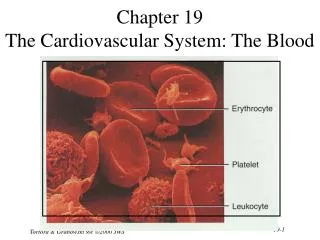
Cardiovascular System – vessels -Chapter 19C. Arteries – Head & Neck. Brachiocephalic trunk – right only – comes directly off the aortic arch

E N D
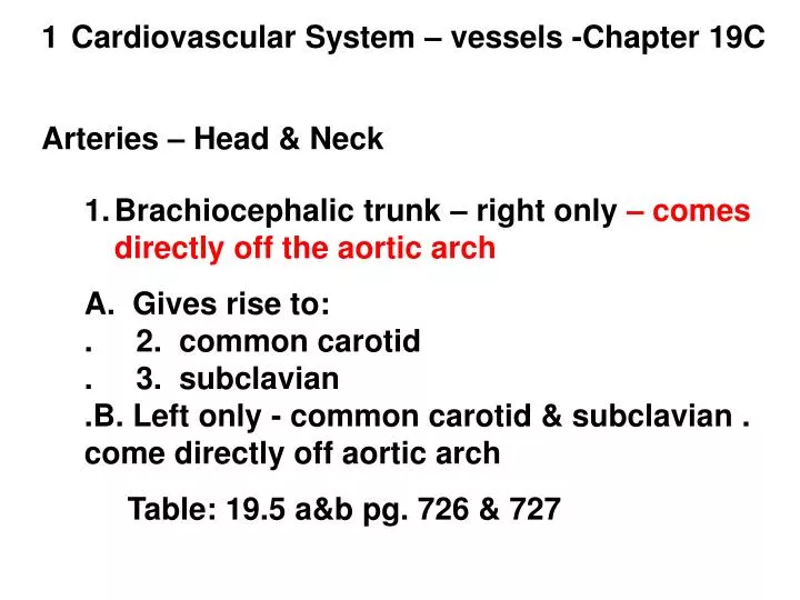
Cardiovascular System – vessels -Chapter 19C Arteries – Head & Neck • Brachiocephalic trunk – right only – comes directly off the aortic arch • A. Gives rise to: . 2. common carotid . 3. subclavian .B. Left only - common carotid & subclavian . come directly off aortic arch • Table: 19.5 a&b pg. 726 & 727
4 C. Both common carotids give rise to: . 4. Internal carotids . 5. External carotids D. Both R & L subclavians give rise to: . 6. L & R vertebral arteries table 19.5 a & b pg. 726 & 727
7 E. Both internal carotids & vertebral arteries . Continue to join and make . 7. Circle of Willis (cerebral arterial circle) Table 19.5 a & c pg 726 & 727
10 Shoulder & Arm - arteries 8. Sub clavians 9. Axiliary 10. Brachial 11. Radial 12. Ulnar Table 19.6 a & b pg. 728 & 729
13 Arteries of the abdomen • Visceral organs of the abdominopelvic cavity • At rest these arteries contain half of all the blood in the body • Branch off the Abdominal aorta 13. Celiac trunk – blood to the stomach, . liver, pancreas, spleen table 19.24 a& b pg. 730 & 731
14 table19.24a pg. 730 Stomach Liver Pancreas spleen
16 14. Superior mesenteric – pancreas, all of the small intestine, most of the large intestine 15. Renal arteries - KIdneys 16. Inferior mesenteric – lower intestine Table 19.7 a, c & d pg. 730 & 733
17 Figure 19.24a, pg. 730 Pancreas All Sm Intestine Most large Lower Intestines
20 Arteries of the pelvis & legs 17. Common iliacs – bifurcation of distal abdominal aorta Common iliacs split to form . 18. Internal iliacs – pelvic viscera & gluteal . Muscles . 19. External iliacs External iliacs leave abdominal cavity and become: 20. femoral arteries, serving the muscles of the thigh: hamstrings, quadraceps & adductors
21. Popiteal: creates an anastomosis in the knee region Popliteal artery bifurcates and forms two branches in the lower leg 22. & 23 anterior & posterior tibial arteries Table 19.8 a & b pgs. 734 & 735
22 Table 19.8a pgs. 734 Commmon iliacs Internal iliacs External iliacs Femoral Popliteal Anterior tibial Posterior tibial
24 Veins – 1. Superior Vena Cava – drains blood from regions superior to the diaphragm 2. L & R brachiocephalics merge to form the superior vena cava Veins draining head and neck Two veins bring blood from the head to the brachiocephalics: 3. Internal jugular 4. Vertebral vein
25 5. Subclavian vein merges w/ brachiocephalic 6. External jugular brings blood from head to the subclavian vein Table: 19.27 a,b,& c pg. 738 & 739
29 Veins that drain blood from hand, wrist & lower arm: 7. Radial 8. Ulnar Ulnar and radial merge and drain blood into: 9. brachial vein which turns into the axillary then subclavian vein 10. Median cuboidal – the vein Dr. takes blood from Table 19.11 a & b pgs: 740 & 741
32 Veins of the abdomen – • Blood drained from the abdominopelvic region enters the inferior vena cava • Veins often follow the paths and share names w/ their arterial counter parts • Veins draining blood from the digestive tract (digested food) all empty into a (12) common hepatic portal vein that goes to the liver to “process” the blood 13 & 14 Superior and inferior mesenteric veins empty into the hepatic portal vein Figure 19.29 pg. 742 & table 19.12 c; pg. 743
35 15. Renal Vein drains the blood from the kidneys taking it to the inferior vena cava Fig. 19.29; pg. 742 Table: 19.12 b page. 743
38 Veins of the pelvis & lower limb • Great and small saphenous veins drain the calf & foot • They are superficial and a common site for varicose veins • Used in coronary artery bi-pass surgery The (16.) anterior tibial and (17) posterior tibial also drain the calf and foot. They join, at the knee and form the knee and form the (18.) popliteal vein. The popliteal continues past the knee and forms the (19.) femoral vein, which drains the thigh, and becomes the (20.) external iliac vein
39The external and (21) internal iliacs join and form the (22) common iliac which drains into the inferior vena cava figure 19.30 a,b & c; page 744