Download
1 / 47
480 likes | 680 Views
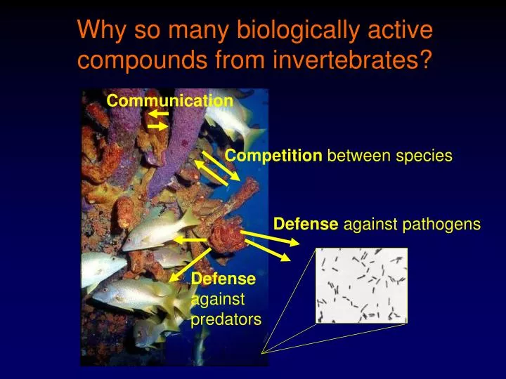
Communication. Competition between species. Defense against pathogens. Defense against predators. Why so many biologically active compounds from invertebrates?. Drugs from the Sea: Invertebrates. Microorganisms. Green Algae. Sponges!!. Brown Algae. Red Algae. Tunicates. Echinoderms.

E N D
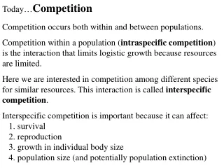
Communication Competition between species Defense against pathogens Defense against predators Why so many biologically active compounds from invertebrates?
Drugs from the Sea: Invertebrates Microorganisms Green Algae Sponges!! Brown Algae Red Algae Tunicates Echinoderms Mollusca Cnidarians (e.g. Corals)
Overview •Introduction to Sponges (Porifera) •Okadaic Acid: Protein Phosphatase Inhibitor •Discodermolide: Potential Anticancer Drug?
Drugs from the Sea: Sponges Out Phylum Porifera > 10,000 species known In Oldest multicellular animal Sessile
Hungry Fish 150,000 bites/m2/day
Yuck! No, thank you! Reject Mmmm! Spongey. Accept Chemical Defenses of Sponges Mix with artificial food Present to fish Extract Percentage (%) Eaten SpongeControl*Treated Acanthella acuta 100.0 6.3 Aplysina aerophoba 89.8 8.2 Ianthella basta 94.0 6.0 Axinella sp. 100.0 93.8 Crambe crambe 94.4 2.8 Stylissa massa 100.0 2.8 Dysidea avara 97.7 27.9 Ircinia fasciculata 100.0 68.9 Petrosia ficiformis 97.5 17.5 * Control = No extract added. Paul and Puglisi (2004), Nat. Prod. Rep., 21:189-209; Paul et al. (2006) Nat. Prod. Rep., 23:153-80.
Bioactive Compounds from Sponges: Okadaic Acid Halichondrin B Okadaic Acid Halichondria okadai
Isolation of Okadaic Acid #1 (Tachibana and Scheuer, Univ. of Hawaii; Van Engen and Clardy, Cornell University) Halichondria okadai 1.MeOH (3x)/Acetone Extraction 2. Remove organic solvent (70% aq.) 3.Hexane Wash (“de-fatting”) 4. EtOAc Extraction Mouse (i.p.) LC50 = 192 µg/kg KB Cytotoxicity 30% Inhibition (2.5 ng/mL) 80% Inhibition (5 ng/mL) Polystyrene Gel, MeOH LH-20, MeOH Si Gel, n-Hexane/Acetone (5:1) Crystallization (from MeOH) Re-Crystallization (from CH2Cl2/Hex.) Colorless Crystalline Solid (0.0001% wet wt.) Tachibana et al. (1981) J. Am. Chem. Soc., 103: 2469-71
Isolation of Okadaic Acid #2 (Gopichand and Schmitz, Univ. of Oklahoma) H. melanodocia 1. 2-Propanol Extraction/H2O dilution 2. CH2Cl2 Extraction 3. 10% MeOH Suspension 4. 10-30% MeOH/Water Suspension 5. Hexane and CCl4 Wash/CHCl3 Ext. Mouse (i.p.) >120 µg/kg Cytotoxicity P388 - ED50 = 1.7 x 10³ L1210 - ED50 - 1.7 x 10² Tumor Inhibition None (≤subtoxic dose) LH-20 (MeOH/CHCl3, 1:1) Silica Gel (CHCl3 to CHCl3 /5% MeOH) Crystallization (from benzene) Crystallization (from benzene/CHCl3) White Crystalline Solid (0.0001% wet wt.) Tachibana et al. (1981) J. Am. Chem. Soc., 103: 2469-71
Okadaic Acid: Structure Elucidation Okadaic Acid MW 804.47 C44H8O13 UV, IR: Uninformative EI-MS: m/z 786 (C44H66O12) 1H and 14C NMR Acetylation (AcO, pyridine, 20 h, r.t.): Tetraacetate (i.e. 4 hydroxyls) Diazomethane Treatment: Methyl Okadaate -> 1H-NMR Comparison to Acanthifolicin: Absolute Stereochemistry Tachibana et al. (1981) J. Am. Chem. Soc., 103: 2469-71
Okadaic Acid: Structure Elucidation Triethyl-Ammonium Okadaate X-Ray Diffraction + o-Bromobenzyl Bromide (in acetone), 36 h (reflux) Si Gel Chromatography Crystallization (2x), CH2Cl2/Hexane o-Bromobenzyl Okadaate
Protein Kinases/Phosphatases: Biochemical “On/Off Switches” ATP ADP Kinase Serine Threonine Tyrosine Phosphatase
Ser/Thr Protein Phosphatases (PP) PP1 PP2A PP4 PP5 PP2B (Calcineurin) PP2C
Ser/Thr Protein Phosphatases 1 and 2A (PP1/2A) PP1 PP2A Catalytic Subunit PP1c(37 Kda) PP2Ac(36 Kda) Distribution Myosin, Glycogen, Widely Chromatin, S.R. Endogenous I-1/DARPP-32, I-2, I-1PP2A, I-2PP2A Inhibitors Dopamine, NIPP-1
Okadaic Acid is a PP1/2A-Specific Inhibitor Phosphatase Substrate ID50 (nM) PP1 PMLC 315 Phosphorylase a 272 PP2Ac PMLC 1.2 Phosphorylase a 1.6 PCM PMLC 205 Phosphorylase a 72 PP2B PMLC 4530 p-Nitrophenyl Phosphate 3600 PP2C PMLC >10,000 Phosphorylase a >10,000 Tyr Phosphatase -- >10,000 Inositol-1,4,5-triPP -- >10,000 Acid Phosphatase -- >10,000 Alkaline -- >10,000 Phosphatase Bialojan and Takai (1988) Biochem. J., 256: 283-90
The “Okadaic Acid Class of Inhibitors” Peptides (+ Nodularins) Microcystins (“Blue-Green Algae”, e.g. Microcystis) Terpenoids Cantharidin (Insects) Thyrsiferyl-23-Acetate (L. obtusa, a “Red Alga”) Other Polyketides Dinophysisotoxin (Dinoflagellate) (+)-Calyculin (Sponge) Tautomycin (Streptomyces)
Discodermolide: Discovery Depth: 33 m Lucaya Discodermia dissoluta
Discodermolide: Isolation Frozen/Thawed 434 g Extracted: MeOH/Toluene (3:1) Partitioned: EtOAc/Water EtOAc Water Column Chromatography (Silica Gel, CH2Cl2/MeOH) Reverse-Phase Chromatography (C18, H2O/MeOH) RP-HPLC (C18, 5µm, 250 x10 mm): 48% H2O/MeOH 7 mg (0.002%) Gunasekara et al. (1990) J. Org. Chem., 55: 4912-4915
Discodermolide: Structure White crystalline solid, mp = 115-6° C UV (MeOH): lmax 235 nm - conjugated dienes IR (CHCl3) : 3600-3500, 1725 cm-1 - hydroxyl and carbonyl Low Resolution FAB-MS:550 Daltons (M+1)+ - CONH2 NMR: 1H, 13C, COSY, HMQC, HMBC NOT Stable at room temperature! Gunasekara et al. (1990) J. Org. Chem., 55: 4912-4915
Discodermolide: Structure 5.0 mg (in 1 mL pyridine) 0.5 mL acetic anhydride (overnight) Acetylation RP-HPLC (C18, 20% H2O/CH3CN) 4.5 mg Gunasekara et al. (1990) J. Org. Chem., 55: 4912-4915
Discodermolide: Structure X-Ray Crystallography
Discodermolide: Synthesis/Structure (+)-Discodermolide (-)-Discodermolide Nerenberg et al. (1993) J. Am. Chem. Soc., 115:12621-2 (and subsequent work by Schreiber Group)
Discodermolide: Synthesis Novartis® Synthesis Scheme
Discodermolide Inhibits Proliferation of Cells Purified Murine (i.e. “mouse”) T-Cell: IC50 = 9 nM Longley et al. (1991) Transplantation, 52: 650-656 Various Human and Murine Cell-Lines: IC50 = 3-80 nM Hung et al. (1994) Chem. Biol., 1:67-71 Estrogen-Receptor Positive/Negative Breast Carcinoma (MCF-7/MDA-MB231): IC50 = 2.4 nM (48 h) Ter Haar et al. (1996) Biochemistry, 35:243-50 NIH3T3 Cells: IC50 Stage (+)-Discodermolide 7 nM (G2/M) (-)-Discodermolide 135 nM (S) Hung et al. (1996) J. Am. Chem. Soc., 118:11054-80
A, T, G, C + DNA Polymerase Mitosis-Promoting Factor (MPF) Cyclin A Cdk2 Cyclin A/B Cdk1 (a.k.a. cdc2) “Restriction Point” Cyclin E Cdk2 G0 Cyclin D Cdk4/6 G0 S G2 Prophase Metaphase G1 Anaphase M Telophase
Microtubules Comprised of Polymers of the Dimer Tubulin b + a -
GTP GTP GTP GTP Tubulin Polymerization Dependent on GTP/GDP Hydrolysis GDP GTP GTP GTP GDP GTP + + GTP GTP GTP GTP
Dynamic Instability of Microtubules Tubulin-GTP Tubulin-GDP
Dynamic Instability of Microtubules Tubulin-GTP Tubulin-GDP
“GTP Cap” Dynamic Instability of Microtubules Tubulin-GTP Tubulin-GDP
Dynamic Instability of Microtubules Tubulin-GTP Tubulin-GDP
Dynamic Instability of Microtubules Tubulin-GTP Tubulin-GDP
Dynamic Instability of Microtubules Tubulin-GTP Tubulin-GDP
Tubulin Polymerization and Depolymerization Aligns Chromosomes During Metaphase Tubulin-Polymerization Dynein - + + - Kinesin Tubulin-Depolymerization
Tubulin Polymerization and Depolymerization Aligns Chromosomes During Metaphase Polymerized Tubulin Dynein - + + - Kinesin
Tubulin Polymerization and Depolymerization Separates Chromosomes During Anaphase Dynein - + + - Tubulin Depolymerizes Tubulin Depolymerizes
Tubulin Polymerization and Depolymerization Separates Chromosomes During Anaphase Dynein - + + -
(+)-Discodermolide Prevents Depolymerization of Tubulin Dynein - + + - Tubulin Depolymerizes Tubulin Depolymerizes
(+)-Discodermolide Stabilizes Microtubules (i.e. Inhibits Depolymerization) Control + Discodermolide
(+)-Discodermolide inhibits depolymerization of tubulin (+)-Discodermolide prevents breakdown of Cyclin B S G2 Mitosis-Promoting Factor (MPF) Cyclin A/B Cdk1 (a.k.a. cdc2) Prophase Metaphase G1 Anaphase M Telophase
Taxol™ (Paclitaxel) * From bark of “Pacific Yew” (Taxus brevifolia)
Discodermolide Stabilizes Microtubules More Than Taxol™ + 10 µM Taxol, or 10 µM (+)-Discodermolide EC50 (+)-Discodermolide 3.2 µM Taxol™ (Paclitaxel) 23 µM
Multi-Drug Resistant Cancer Cells Less Resistant to Discodermolide “Level of Resistance”* Colon Ovarian CarcinomaCarcinoma (+)-Discodermolide 25-fold 89-fold Taxol 900-fold 2800-fold *Compared to parent line
(+)-Discodermolide Binds to Same (or Overlapping Site) as Taxol
FDA Drug Approval: An Overview Discovery Pre-Clinical Toxicity/Pharmacology in vitro and in vivo (animal models, e.g. rodents) How much of the drug is absorbed in the blood? How is the drug broken down in the body? What is the toxicity of the drug and its breakdown products? How quickly does the body excrete the drug and its by-products? Synthesis and/or Purification Clinical Trials Phase 1: 20-80 patients; safety, safe dose, side-effects Phase 2: 40-100 patients; effectiveness, further safety Phase 3: 200+ patients; effectiveness, comparison, further safety Phase 4: After drug marketed; safety in particular groups, long-term effects