Download
1 / 65
660 likes | 812 Views
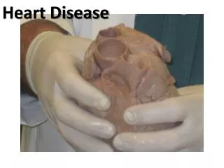
Acquired Heart Disease. Prof. Abdullah Al-Jarallah, MD. Pediatric Cardiologist. Acquired Heart disease. Disease affecting cardiac tissue and function which does not have its inception at birth and usually is secondary to an extraneous agent. Types of Acquired Heart Disease.

E N D
Acquired Heart Disease Prof. Abdullah Al-Jarallah, MD. Pediatric Cardiologist
Acquired Heart disease Disease affecting cardiac tissue and function which does not have its inception at birth and usually is secondary to an extraneous agent.
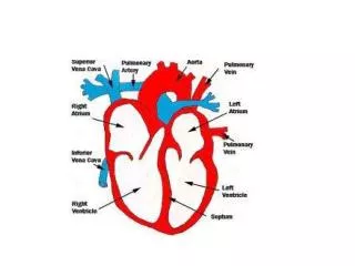
Types of Acquired Heart Disease Ischemic / hypoxic Hypertensive infectious Inflammatory Metabolic Nutritional Traumatic
Ischemic / Hypoxic Coronary occlusion Atherosclerosis Kawasaki Disease Sickle cell anemia Hypoperfusion Surgical ischemic arrest severe hypotension Asphyxia
Hypertensive Systemic hypertension Pulmonary hypertension
Infectious Pericarditis Myocarditis Endocarditis
Inflammatory Post-pericardiotomy Syndrome Rheumatic fever Collagen vascular Diseases
Metabolic Endocrine adenopathy Adrenal Pituitary Pancreatic Thyroid Parathyroid Storage diseases Glycogen Mucopolysacchrides
Nutritional Nutritional deficiencies Starvation Vitamin Mineral Carnitine Nutritional Excesses Obesity Vitamin Mineral
Traumatic Penetrating Blunt
Kawasaki Disease Pericarditis and post-pericardiotomy syndrome Myocarditis / congestive Cardiomyopathy Infectious Endocarditis Rheumatic heart disease
Kawasaki Disease Recognized in 1970’s Inflammatory disease of unknown etiology 9.2/100,000 cases per year; usually <4 y/o Winter and spring; 3yr”epidemics” Asiatics and blacks > white: 9&1.5/1
Kawasaki’s Disease Pathophysiology Immunoregulatory anomalies Activation of T and B lymphocytes Production of immunoglobulins and cytokines wide spread immune reaction Generalized microvasculitis Myocardial and pericardial inflammation Coronary vasculitis
Kawasaki’s Disease Clinical Manifestations Fever of 5 + days duration Physical findings Polymorphous rash Non-purulent conjunctivitis Erythema of oral membranes including tongue Indurative edema of hands and feet Cervical lymphadenopathy Acute and often severe toxic presentation multi-organ involvement
Kawasaki’s Disease Laboratory Findings Elevated acute phase reactants Elevated ESR Elevated Platelets Myocardial dysfunction Pericardial effusion Coronary thickening ---> coronary dilatation and aneurysm
Kawasaki's Disease- Cardiovascular Stages 20% of untreated; 2-4% with treatment Stage1: Week 1-2 Microvasculitis Peri, myo, and endocarditis endocarditis and perivasculitis of coronaries Stage 2: Week 1.5-3 Vasculitis of coronaries with aneurysms and thrombi intimal proliferation of coronaries peri, myo-, and endocarditis Stage 3: Week 4-5 Scaring and intimal thickening of Coronaries Myocardial infarction Stage 4: >2m0 Advanced coronary artery disease Myocardial Fibrosis
Echocardiographic Findings Acute phase: Pericardial effusion LV dysfunction Diffuse coronary artery wall thickening and dilatation in 30-50% Coronary dilatation <5y/o, lumen >3 mm Sacular or fusiform
Treatment IVGG: 2g/kg over 24 hrs ASA: 20-25 mg/kg/dose, q 6 hrs until afebrile 2-3 days 3-5 mg/kg/day 6-8 weeks, until ESR and plt count normal Indefinitely if coronary artery anomalies
Pericarditis / post-pericardiotomy Syndrome Inflammation(infection) of pericardial space Chest pain Friction rub Pericardial effusion Fever Elevated ESR
Pericarditis Viral Purulent Tuberculous Rheumatic Kawasaki Uremic
Post - pericardiotomy Syndrome 30%, if pericardium opened 1-2 weeks post surgery Etiology?? Viral Autoimmune Symptoms: Fever Chest pain Friction rub Pericardial effusion
Post - pericardiotomy syndrome Treatment: ASA: 50-75 mg/kg/day; 4-6 weeks Steroids: 2mg/kg/day; taper over 3-4 weeks Diuretics (cautiously)
Cardiac tamponade Pathophysiology Increase in pericardial fluid which elevates filling pressures, impedes ventricular filling and decreases cardiac output Rapid small volume increase versus large chronic volume
Cardiac Tamponade Physical findings Decreased heart sounds Distended jugular veins Pulsus paradoxus >10 mmHg decrease in SBP with inspiration Increased pooling of blood in pulmonary bed due to decreased LV filling
Cardiac tamponade ECG: Low voltage ST - T wave changes Electrical alternans CXR “Water - bottle” heart, if large volume Normal, if acute ECHO space between heart and pericardium Swinging heart Inspiratory variation in Doppler flows
Myocarditis / Congestive Cardiomyopathy Infection of myocardium with lymphocytic infiltration Degenerative process affecting myocytes Impairment of myocardial function
Myocarditis - Etiology Viral - Coxackievirus, ECHO, adeno, etc. Bacterial - Tuberculosis, strep, etc. Fungal - unusual Protozoan - Chaga’s disease (T.cruzi), malaria, toxoplasmosis Rickettsial Spirochetal metazoal - trichinosis, echinococcosis, etc.
Congestive Cardiomyopathy - Etiology Infectious - viral familial - duchenne’s Metabolic - glycogen storage Ischemic - Kawasaki Toxic - anthracyclines Nutritional - carnitine
Clinical Manifestation General malaise or viral syndrome low cardiac output state (Shock) Gallop rhythm (mitral insufficiency) ST - T wave changes ECHO: Reduced shortening fraction Segmental wall motion anomalies Valvar insufficiency
Course Myocarditis 70-80% 20-30% Mild-Mod CHF Severe CHF 60-70% 10-20% 10% Recovery Dil.Cardiomyo Death/Trans
Diagnosis Clinical findings Identification of etiologic agent Endomyocardial biopsy Lymphocytic infiltrate Etiologic agent Necrosis “Staging”
Treatment Symptomatic inotropes Diuretics afterload reduction Correction of etiology Immunosupression Steroids Anti-virals Cyclosporin Interferon Transplantation
Infective Endocarditis Microbial infection of endocardial surface of heart - valves or wall “Acute” (virulent) / “subacute” (prolonged) 1:1800 to 1:4500 ped cases: admissions Any age; greater in 5th decade Pre-v. post-antibiotic era - no change Factors: Better diagnosis Drug abuse Treatment modalities
Etiology Alpha hemolytic strep: most common (>60%); prolonged Staph aureus: 2nd most common (20%); virulent Beta hemolytic strep: uncommon Coagulase negative Staph: increasing Candida
Risk Factors High Risk Prosthetic valves Surgical shunts Indwelling catheters Previous SBE Moderate Risk PDA VSD ASD (not secondum) Bicuspid aortic valve RHD MVP with MR
Clinical Manifestations Fever High (Staph) Low (Strep) “Viral syndrome” New murmur CHF Petechiae Inc ESR, anemia, hematuria
Diagnosis Blood Culture Positive off antibiotics 5-8% negative cultures 2-3 sets over 24 hrs; (as much as possible) ECHO: Vegitations Valve insufficiency
Treatment Specific anti-microbial 4-6 weeks IV 2 weeks w/wo P.O. Surgery, esp. prosthetic valves
Prophylaxis AHA guidelines Amoxicillin - oral, upper resp procedures Clindamycin (penicillin allergic) Amp and gent or vancomycin - GU or GI
Rheumatic Fever Most common cause of acquired heart disease in children (5-15 y peak of 8 y) USA: 0.5-3.0/100,000 (1900: 100-200/100,000) Post- infectious connective tissue response in susceptible host Group A beta- hemolytic streptococcus infection of the pharynx F/H of RHD and low socioecnomic status.