Download

1 / 83

E N D
ENDOTHELIAL CELLS • ECs comprise the single cell-thick, continuous lining of the entire cardiovascular system, collectively called the endothelium. Endothelial structural and functional integrity is fundamental to the maintenance of vessel wall homeostasis and normal circulatory function.
Smooth muscle cells • SMCs are predominant cellular element of the vascular media • SMCs are responsible for vasoconstriction and dilation in response to normal or pharmacologic stimuli. • They also synthesize collagen, elastin, and proteoglycans; and elaborate growth factors and cytokines. They migrate to the intima and proliferate following vascular injury. • Thus, SMCs are important elements of both normal vascular repair and pathologic processes such as atherosclerosis.
Smooth muscle cells • Vascular injury ( endothelial injury/dysfunction) stimulates SMC growth. Reconstitution of the damaged vascular wall is a physiologic healing response that includes the formation of a neointima, in which SMCs (1) migrate from the media to the intima, (2) multiply as intimal SMCs, and (3) synthesize and deposit ECM
Smooth muscle cells • During the healing response, SMCs undergo changes that resemble dedifferentiation. In the intima they lose the capacity to contract and gain the capacity to divide. • Intimal SMCs may return to a nonproliferative state when either the overlying endothelial layer is re-established following acute injury or the chronic stimulation ceases.
Arteriosclerosis Arteriosclerosis (literally, "hardening of the arteries") is a generic term for thickening and loss of elasticity of arterial walls. Three patterns of arteriosclerosis are recognized; they vary in pathophysiology and clinical and pathological consequences. 1)Atherosclerosis, the most frequent and important pattern
Arteriosclerosis 2)Mönckeberg medial calcific sclerosis is characterized by calcific deposits in muscular arteries in persons older than age 50. They do not encroach on the vessel lumen. 3)Arteriolosclerosis affects small arteries and arterioles. There are two anatomic variants, hyaline and hyperplastic, both associated with thickening of vessel walls with luminal narrowing that may cause ischemic injury. Most often associated with hypertension and diabetes mellitus.
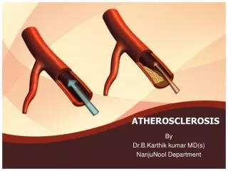
Atherosclerosis • Atherosclerosis is characterized by intimal lesions called atheromas, or atheromatous or fibrofatty plaques, which protrude into and obstruct vascular lumens and weaken the underlying media. They may lead to serious complications
Atherosclerosis: Morphology • Fatty streaks are the earliest lesion of atherosclerosis. They are composed of lipid-filled foam cells. They are not significantly raised and thus do not cause any disturbance in blood flow. Fatty streaks begin as multiple yellow, flat spots less than 1 mm in diameter that coalesce into elongated streaks, 1 cm long or longer. They contain T lymphocytes and extracellular lipid in smaller amounts than in plaques.
Fatty streak—a collection of foam cells in the intima. A. Aorta with fatty streaks ( arrows), associated largely with the ostia of branch vessels. B. Close-up photograph of fatty streaks from the aorta of an experimental hypercholesterolemic rabbit shown after staining with Sudan red, a lipid-soluble dye, again illustrating the relationship of the lesions to the two-branch vessel ostia. C. Photomicrograph of fatty streak in an experimental hypercholesterolemic rabbit, demonstrating intimal macrophage-derived foam cells ( arrow). Slide 12.9
Morphology • The key processes in atherosclerosis are intimal thickening and lipid accumulation. An atheroma or atheromatous plaque consists of a raised focal lesion initiating within the intima, having a soft, yellow, grumous core of lipid (mainly cholesterol and cholesterol esters), covered by a firm, white fibrous cap.
Morphology • The atheromatous plaques appear white to whitish yellow and impinge on the lumen of the artery. They vary in size from approximately 0.3 to 1.5 cm in diameter but sometimes coalesce to form larger masses. Atherosclerotic lesions usually involve only a partial circumference of the arterial wall ("eccentric" lesions) and are patchy and variable along the vessel length.
Atherosclerosis: Morphology • The most heavily involved vessels are the abdominal aorta then coronary arteries, the popliteal arteries, the internal carotid arteries, and the vessels of the circle of Willis.
Atherosclerosis: Morphology Atherosclerotic plaques have three principal components: • (1) cells, including SMCs, macrophages, and other leukocytes • (2) ECM, including collagen, elastic fibers, and proteoglycans • (3) intracellular and extracellular lipid . These components occur in varying proportions.
Atherosclerosis: Morphology • Typically, the superficial fibrous cap is composed of SMCs and relatively dense ECM. Beneath and to the side of the cap (the "shoulder") is a cellular area consisting of macrophages, SMCs, and T lymphocytes. • Deep to the fibrous cap is a necrotic core, containing a disorganized mass of lipid (primarily cholesterol and cholesterol esters), cholesterol clefts, debris from dead cells, foam cells, fibrin, variably organized thrombus, and other plasma proteins.
Atherosclerosis: Morphology • Foam cells are large, lipid-laden cells that derive predominantly from blood monocytes (tissue macrophages), but SMCs can also imbibe lipid to become foam cells. • Around the periphery of the lesions, there is usually evidence of neovascularization (proliferating small blood vessels). Typical atheromas contain relatively abundant lipid. • Atheromas often undergo calcification.
Major components of well-developed atheromatous plaque: fibrous cap composed of proliferating smooth muscle cells, macrophages, lymphocytes, foam cells, and extracellular matrix. The necrotic core consists of cellular debris, extracellular lipid with cholesterol crystals, and foamy macrophages. Slide 12.6
Gross views of atherosclerosis in the aorta. A. Mild atherosclerosis composed of fibrous plaques, one of which is denoted by the arrow. B. Severe disease with diffuse and complicated lesions. Slide 12.7
Histologic features of atheromatous plaque in the coronary artery. A. Overall architecture demonstrating a fibrous cap (F) and a central lipid core (C) with typical cholesterol clefts. The lumen (L) has been moderately narrowed. Note the plaque-free segment of the wall ( arrow). In this section, collagen has been stained blue (Masson trichrome stain). B. Higher-power photograph of a section of the plaque shown in A, stained for elastin ( black) demonstrating that the internal and external elastic membranes are destroyed and the media of the artery is thinned under the most advanced plaque ( arrow). C. Higher-magnification photomicrograph at the junction of the fibrous cap and core showing scattered inflammatory cells, calcification ( broad arrow), and neovascularization ( small arrows). Slide 12.8
American Heart Association classification of human atherosclerotic lesions from the fatty dot (type I) to the complicated type VI lesion. The diagram also includes growth mechanisms and clinical correlations. Slide 12.11
COMPLICATIONS The advanced lesion of atherosclerosis is at risk for the following pathological changes that have clinical significance: 1) Focal rupture, ulceration, or erosion of the luminal surface of atheromatous plaques may result in exposure of highly thrombogenic substances that induce thrombus formation or discharge of debris into the bloodstream, producing microemboli composed of lesion contents (cholesterol emboli or atheroemboli).
COMPLICATIONS 2) Hemorrhage into a plaque, especially in the coronary arteries, may be initiated by rupture of either the overlying fibrous cap or the thin-walled capillaries that vascularize the plaque. A contained hematoma may expand the plaque or induce plaque rupture.
COMPLICATIONS 3)Superimposed thrombosis, the most feared complication, usually occurs on disrupted lesions (those with rupture, ulceration, erosion, or hemorrhage) and may partially or completely occlude the lumen. Thrombi may heal and become incorporated into and thereby enlarge the intimal plaque.
4)Aneurysmal dilation may result from ATH-induced atrophy of the underlying media, with loss of elastic tissue, causing weakness and potential rupture 5) Calcifications.
Natural history of atherosclerosis Slide 12.5
Risk Factors for Atherosclerosis Major Nonmodifiable • Increasing age • Male gender • Family history • Genetic abnormalities PotentiallyControllable • Hyperlipidemia • Hypertension • Cigarette smoking • Diabetes
Risk Factors for Atherosclerosis Lesser, Uncertain, or Nonquantitated • Obesity • Physical inactivity • Stress ("type A" personality) • Postmenopausal estrogen deficiency • High carbohydrate intake • Alcohol • Lipoprotein Lp(a) • Hardened (trans)unsaturated fat intake • Chlamydia pneumoniae
Types of lipoproteins • Low-density lipoproteins (LDLs): When too much LDL (bad) cholesterol circulates in the blood, it promotes atheroma formation in the arteries.LDLs contribute to heart disease because they carry large amounts of cholesterol. • Very-low-density lipoproteins (VLDLs): is also considered to be a type of bad cholesterol because it helps cholesterol build up on the walls of arteries • Chylomicrons also promote atherosclerosis.
Types of lipoproteins • High-density lipoproteins (HDLs): is known as “good” cholesterol, because high levels of HDL seem to protect against heart attack. Low levels of HDL (less than 40 mg/dL) also increase the risk of heart disease. HDLs help to reverse the effects of high cholesterol by collecting cholesterol from other lipoproteins and transporting it to places where it can be utilized by the cells
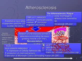
PATHOGENESIS: response to injury hypothesis • This concept, called theresponse to injury hypothesis, considers atherosclerosis to be a chronic inflammatory response of the arterial wall initiated by injury to the endothelium. Moreover, lesion progression is sustained by interaction between modified lipoproteins, monocyte-derived macrophages, T lymphocytes, and the normal cellular constituents of the arterial wall.
Processes in the response to injury hypothesis. 1, Normal. Slide 12.13
2, Endothelial injury with adhesion of monocytes and platelets (the latter to denuded endothelium). Slide 12.14
3, Migration of monocytes (from the lumen) and smooth muscle cells (from the media) into the intima. Slide 12.15
4, Smooth muscle cell proliferation in the intima. Slide 12.16
5, Well-developed plaque. Slide 12.17
PATHOGENESIS: response to injury hypothesis Central to this thesis are the following: • Accumulation of lipoproteins, mainly LDL, with its high cholesterol content, in the vessel wall • Chronic endothelial injury, usually subtle, • increased permeability, leukocyte adhesion, and thrombotic potential.
PATHOGENESIS: response to injury hypothesis • Adhesion of blood monocytes (and other leukocytes) to the endothelium, followed by their migration into the intima and their transformation into macrophages and foamcells • Adhesion of platelets • Release of factors from activated platelets, macrophages, or vascular cells that cause migration of SMCs from media into the intima
PATHOGENESIS: response to injury hypothesis • Proliferation of smooth muscle cells in the intima, and elaboration of extracellular matrix, leading to the accumulation of collagen and proteoglycans • Enhanced accumulation of lipids both within cells (macrophages and SMCs) and extracellularly.
PREVENTION • primary prevention programs, aimed at either delaying atheroma formation or causing regression of established lesions in persons who have never suffered a serious complication of atherosclerotic coronary artery disease • secondary prevention programs, intended to prevent recurrence of events such as myocardial infarction in patients with symptomatic disease.
PREVENTION • based on risk factor modification: abstention from or cessation of cigarette smoking, control of hypertension, weight reduction and increased exercise, moderation of alcohol consumption, and, most importantly, lowering total and LDL blood cholesterol levels while increasing HDL. on
Rheumatic Fever and Heart Disease • Definition: rheumatic fever is an acute, immunologically mediated, multi-system inflammatory disease that follows, after a few weeks, an episode of group A beta hemolytic streptococcal pharyngitis (3% of patients). • The incidence and mortality of rheumatic fever has declined over the past 30 years (due to improved socioeconomic condition and rapid diagnosis and treatment of strep. pharyngitis.
Rheumatic Fever: Heart • Affect the heart during its acute phase acute rheumatic carditis. • Cause chronic valvular deformities (many years after the acute disease.
Pathogenesis and Key Morphologic Changes of Acute Rheumatic Heart Disease Hypersensitivity reaction induced by group A strept. (ab. Against protein M Cross-reaction / Autoimmune response
Morphology: Acute Rheumatic Fever • Inflammatory infiltrates occur in a wide range of tissues: synovium, joints, skin, heart. • Focal fibrinoid necrosis mixed inflammatory reaction (diffuse or localized) Fibrosis (chronic rheumatic heart disease) .
Acute Rheumatic Carditis • Pancarditis (endo- myo- pericarditis). • Multiple foci of inflammation within the connective tissue of the heart. (Aschoff bodies: central fibrinoid necrosis, surrounded by chronic mononuclear inflammatory infiltrate and occasional large histiocytes). • Diffuse interstitial inflammatory infiltrates (may lead to generalized dilation of the cardiac chambers).
Acute Rheumatic Carditis • Pericardial involvement: fibrinouspericarditis, sometime associated with serous or serosanguinous effusion. • Endocardium: • Mostly mitral and aortic valve. • Valves are edematous and thickened with foci of fibrinoid necrosis. (Aschoff nodules uncommon). • Verrucousendocarditis (small vegetations along lines of valve closure). • Acute changes may resolve completely or progress to scarring and chronic valvular deformities.