Download
1 / 35
400 likes | 724 Views
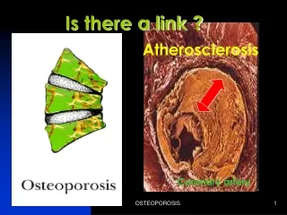
ATHEROSCLEROSIS. Definition: Atheroma = porridge. Atherosclerosis is defined as formation of atheromas or atheromatous or fibrofatty plaques in the intima of large and medium sized muscular arteries producing narrowing of the lumen and weakening of the media. EPIDEMIOLOGY.

E N D
ATHEROSCLEROSIS Definition: Atheroma = porridge. Atherosclerosis is defined as formation of atheromas or atheromatous or fibrofatty plaques in the intima of large and medium sized muscular arteries producing narrowing of the lumen and weakening of the media.
EPIDEMIOLOGY Highest incidence in developed countries. Highest mortality due to IHD. Risk Factors: Non-modifiable: Major: Age, male sex, family history, genetic abnormalities. Minor: Obesity, sedentary activity, stress – type A personality. Modifiable: Major: Hyperlipidemia, hypertension, diabetes, cigarette smoking. Minor: Alcoholism, infections, homocystinuria, saturated fat intake
Major Non-Modifiable Risk Factors Age: Dominant influence. Risk increase with each decade. Sex: Males at higher risk of developing atherosclerosis and its complication. Females are protected by estrogen. Genetic basis: Familial tendency as with familial hypercholesterolemia, hypertension and diabetes; polygenic.
Major Modifiable Risk Factors Hypercholesterolemia: Sufficient for the development of atherosclerosis in the absence of other risk factors. Increased risk with increased levels of LDL (bad cholesterol) that delivers cholesterol to the periphery. HDL mobilizes cholesterol from atheromas and transports to liver for its excretion in bile. Higher the levels of HDL lower the risk
Exercise and moderate alcohol intake; increase HDL level. Obesity and smoking decrease HDL levels. High dietary intake of saturated fat; egg yolk, animal fat, butter raises plasma cholesterol. Polyunsaturated vegetable oils; Polyunsaturated fat, omega-3 fat in fish; beneficial
Hypertension: Major risk factor. Both systolic and diastolic blood pressure associated with increased risk. Cigarette smoking: Both in men and women. More the number more is the risk. Cessation; decreases the risk. Diabetes: Induces hypercholesterolemia. Increased predisposition to atherosclerosis. Other Factors Higher risk: High homocyteine levels. High Lp(a) levels. Type A personality. Sedentary activity. Obesity. Lower risk: Moderate alcohol intake
Multiple risk factors, multiplicative effect. 2 major risk factor = 4 times the risk, 3 major risk factor = 7 times the risk. Atherosclerosis and its complication in an individual in the absence of any apparent risk.
Fatty Streaks The earliest lesions of atherosclerosis. Composed of lipid filled foam cells. Do not cause any disturbance to the blood flow as the lesions are not significantly raised form the surface. Start as yellow multiple spots less than 2mm and join to form elongated streaks of 1cm or more. In aorta they are seen in all children 10 years or more regardless of any sex, race, geography and environment. In coronary artery starts in adolescence. Precursors of atheromatous plaque.
Distribution: More common in abdominal aorta and the lesions are more prominent around the ostia of the major branches. Order of frequency: Abdominal aorta, coronary arteries, popliteal arteries, internal carotid arteries and vessels of circle of Willis. Vessels of upper extremities are spared. Mesenteric and renal artery ostia.
Atheromatous Plaque: Atheromatous plaque primarily develop in, Elastic arteries – aorta, carotid and iliac arteries. Large and medium sized muscular arteries – coronary and popliteal arteries. Symptomatic plaques are seen in coronary artery, carotid artery, renal artery, arteries of lower extremities.
Morphology: Atheromatous plaque also called as fibrous, fibrofatty, lipid or fibrolipid plaques. White or whitish yellow. Raised on the surface obstructing the lumen. 0.3 to 1.5 cm but may be larger. Involve only the partial circumference
Morphology: Patchy; along the length of the vessel. With progression; number increases and become diffuse. Consists of; Raised focal lesion. A soft yellow central lipid core. A firm white fibrous cap covering the central core.
Microscopy: Three components: Cells: Smooth muscle cells, macrophages and other leucocytes. Extracellular matrix: Collagen, elastic fibers and proteoglycans. Intracellular and extracellular lipid. Fibrous cap; smooth muscle cells and dense extracellular matrix. Beneath and at the sides of cap (shoulder); macrophages, smooth muscle cells and T lymphocytes. Deep to fibrous cap; central core, composed of necrotic debris, mass of lipid, cholesterol clefts, foam cells and fibrin. Periphery of the lesion; neovascularization
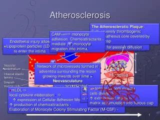
Pathogenesis Role of Endothelial Injury Chronic and repetitive endothelial injury is the important predisposing factor in the pathogenesis of atherosclerosis. In the absence of endothelial injury endothelial dysfunction; causes increased endothelial permeability and atherogenecity. Factors that influence endothelial dysfunction are derivatives of cigarette smoke, homocysteine, infectious agent and cytokines.
ROLE OF INFLAMMATION Inflammatory mechanism mediate initiation, progression and complication of atherosclerosis. In early lesions the endothelial cells express adhesion molecules that binds to leucocytes especially monocytes and T lymphocytes. Monocytes adhere to endothelium, migrate to intima, transform into macrophages, engulf lipoproteins and transform into foam cells.
Foam Cells: Derived from blood monocytes and tissue macrophages. Large lipid laden cells.
Macrophages release growth factors and cytokines causing migration of leucocytes and proliferation of smooth muscle cells. T lymphocytes and macrophages mediate chronic inflammation and deposition of extracellular matrix that is the characteristic feature of late atheromatous plaque.
ROLE OF LIPIDS Mechanism of hyperlipidemia causing atherosclerosis: Chronic hypercholesterolemia causes endothelial dysfunction by increased production of oxygen free radicals that deactivate NO, endothelial relaxing factor. Accumulated lipid is oxidized and ingested by macrophages that increase the monocyte accumulation, stimulate CK and GF that are toxic for endothelial cells and ECM.
ROLE OF LIPIDS Dyslipoproteinemias result from either Mutation that yield defective apolipoprotein Other disorders like nephrotic syndrome, alcoholism, hypothyroidism or diabetes mellitus. Abnormal lipoprotein patterns are increased LDL cholesterol, decreased HDL cholesterol and increased abnormal Lp(a).
Role of Smooth Muscle Cells Smooth muscle cells migrate from media to intima, proliferate, deposit extracellular matrix and convert fatty streak into a atheromatous plaque. Growth factors that are responsible for smooth muscle cell proliferation are PDGF, FGF and TGF. Other Factors Oligoclonal lesions: Equivalent to benign neoplasm induced by exogenous chemical or oncogenic viruses. Infection: Chlamydia pneumoniae and cytomegalovirus. Potentiate chronic inflammatory response and hypercholesterolemia.
Complication of Atheromatous Plaque: Rupture, ulceration and erosion: Exposes the highly thrombogenic substances leading to thrombus formation. Atheroemboli: Also called as cholesterol emboli. Discharge of fat debris into the circulation after the rupture of the plaque.
Hemorrhage into the plaque: Rupture of fibrous cap or blood vessels; hematoma. Thrombosis: Most dreaded complication. On a disrupted lesion. Aneurysm formation: Due to atrophy of media and loss of elastic fibers; wall weakening and permanent dilatation.
Major Consequences: Myocardial infarction. Cerebrovascular accidents (stroke). Aortic aneurysm. Lower limb gangrene. Mesenteric occlusion
Clinical Effects Aneurysm and rupture due to weakening of the media. Occlusion due to thrombosis. Critical stenosis leading to ischemia and infarction of the affected tissue or organ.
Prevention By risk factor modification. Primary prevention: Aimed to delay the atheroma formation or regressing the established lesions in patients who never suffered serious complications. Secondary prevention: Aimed to prevent the recurrence of complications of atherosclerosis like MI. cessation of cigarette smoking. Control of hypertension. Weight reduction. Increasing exercise. Moderate alcohol consumption. Lowering total and LDL cholesterol and increasing HDL cholesterol.