Download

1 / 43
540 likes | 1.63k Views
Pulmonary Embolism Aortic Aneurysm Aortic Dissection. Nursing 313, Fall 2011. Incidence. >650,000 cases diagnosed per year in US Third most common cause of death in hospitalized patients Greatest risk those who have a DVT Recent trends: men > women Risk doubles every ten years after 60.

E N D
Pulmonary EmbolismAortic AneurysmAortic Dissection Nursing 313, Fall 2011
Incidence • >650,000 cases diagnosed per year in US • Third most common cause of death in hospitalized patients • Greatest risk those who have a DVT • Recent trends: men > women • Risk doubles every ten years after 60
Risk Factors http://www.cholesterolcholestrol.com/virchows-triad.jpg
Pathophysiology Clot lodges in PA or branches ↑ in Dead Space → VQ Mismatch Release of Vasoactive Substances ↑ in Vaso & Bronchoconstriction ↑ PA Pressures ↑ RV workload → RV Failure
Respiratory System • A balancing act between ventilation (V) and perfusion (Q)
Perfusion / ventilation mismatch V/Q mismatch • Obstructed area has absent or diminished blood flow • Alveoli ventilated but not perfused which causes increased dead space • Severity depends on size of embolism and degree of vascular obstruction • Possibility of severe hypotension and shock
Signs and Symptoms • Sudden onset of dyspnea • Sudden onset of pleuritic chest pain • General signs of hypoxemia • Feeling of impending doom • ↑ Anxiety • Cough, possible hemoptysis • Mild fever • Diaphoresis
If severe, then cardiac involvement • Cardiac s/s are due to loss of the forward flow of blood • JVD • S3 or S4 • Syncope • Tachycardia • Tachypnea • Rales • Signs of right sided heart failure
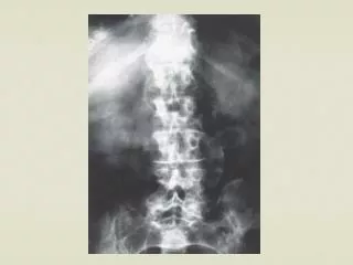
Diagnosis • D-Dimer assay – possible method of ruling out a PE • Spiral CT scan • Pulmonary Angiography (gold standard) • V/Q scan • CXR, ECG, ABGs
Emergency Management • Stabilize the cardiopulmonary system • Oxygen • Insertions of IV lines • Treat hypotension – vasoactive drugs if ↓BP • Diagnostic testing: scans, ABGs, labs • Indwelling urinary catheter • Sedatives or pain relief as needed • Prevent further emboli from forming
Pharmacological Management Heparin • Heparin- initial and preferred treatment • Does not affect the existing clot • PTT/ therapeutic range 1.5 to 2 times normal • Antidote: protamine sulfate
Warfarin • Warfarin (Coumadin) • Interferes with the synthesis of the vitamin K • 3-4 days for therapeutic benefit • INR between 2.0 – 2.5 for those with PE • Antidote – Vitamin K
Thrombolytic Therapy • tPA, Alteplase, Reteplase • Converts plasminogen to plasmin • Specific contraindications: • Recent CVA; active bleeding; surgery in past 10 days; trauma; recent labor and delivery, severe hypertension • Major complication: bleeding
Nursing Management of Patients on Anticoagulation Therapy • Frequent vital signs • Hematest stools • Handle patient gently • Avoid IM injections & veni-punctures • Use electric shaver & soft toothbrushes • Nothing per rectum • Avoid foods containing vitamin K • Monitor labs
Surgical Management • Thrombectomy or Embolectomy • Vena Cava Interruption (Filters) • Indicated in patients who may not tolerate anticoagulation
Nursing Management • Identify patients at risk • Manage pain! • Psychosocial support for patient and family • Utilize nursing interventions to minimize risk • Early ambulation • Watch labs, VS, targeted assessments for cardiopulmonary system • Patient & family teaching on importance of lifestyle changes
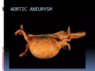
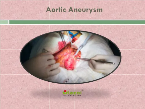
Factoids • Atherosclerosis damages the lining of the aorta • Majority below the renal arteries • Exact cause of aneurysm unknown • More common in Caucasians • More common in men than women • Higher risk of rupture and death in women compared with men with same size aneurysms • Rare in young females, but related to pregnancy
Incidence & Risk Factors • 13th leading cause of death in men aged 65-75 • Up to 13% of individuals with AA have multiple aneurysms • Causes about 9,000 deaths / year • Risk factors: • Atherosclerosis • Patients born with bicuspid aortic valves (new) • Hypertension • Age • Smoking, cholesterol, ↑lipids • Genetic CT disorders (Marfan’s) • History of crack use in pregnancy • Positive family history • Often found incidentally
Classification: Location • Abdominal • More common (75%) • Usually infra-renal • Thoracic • Less common (25%) • > chance of rupture & dissection
Clinical Manifestations • Abdominal Aortic Aneurysms • Asymptomatic (early) • Symptoms caused by expansion (later) • Chest or abdominal pain, flank pain, scrotal pain • Pulsatile mass palpated (80%) , bruit + • Feels like heart beating in abdomen • Ruptured aneurysm is a medical emergency
Clinical Manifestations Thoracic Aortic Aneurysms • Asymptomatic (early) • Symptoms by compression of tissue (later) • Ascending: CHF due to aortic regurgitation, edema of the UE and face • Arch or descending: wheeze, cough, hemoptysis, dysphagia, hoarseness • Chest or back pain is common to both
Aortic Dissection • Occurs when there is a tear in the intimal lining (or adventitia) and blood gets diverted into the channel → ↓ intravascular volume • http://www.youtube.com/watch?v=ZtanUq95pTk
More common in thoracic aneurysms May interfere with major aortic branches causing organ failure Classification system: Debakey Stanford Clinical manifestation: PAIN!!!!
Aortic Dissection Classifications • DeBakey Classification: • Type I - originated in ascending aorta • Type II - originated in and is confined to ascending aorta • Type III - originated in descending aorta • Stanford Classification • A - Originated and involves ascending aorta. • B - Originated and involves descending aorta
Dx tests for Aortic Dissections • Trans thoracic echocardiogram (TTE) • Trans esophageal echocardiogram (TEE) • Computed Tomography Angiography (CTA) • Magnetic Resonance Angiography • Ultrasonography • Aortography
Medical Management ofAortic Aneurysm • Control /eliminate risk factors • F/U every 3 to 6 months (ultrasounds) • Medications • Beta-blockers/calcium channel agents (older) • Recent evidence favors ACE inhibitors over BB • Statins, Doxycycline: both inhibit matrix metalloproteinases (MMPS) • MMPs are enzymes that break down elastin and collagen in the aortic wall – contributes to aneurysm formation
General Indications for Surgical Repair of Aortic Aneurysms • Diameter ≥5.5 cm (men) • For women, 4.5-5.0 cm (due to greater incidence of rupture) • Ascending Thoracic • Diameter ≥5.5 cm (5 cm in patients with Marfan syndrome) • Symptoms suggesting expansion or compression of surrounding structures • Rapidly expanding aneurysms (growth rate >0.5 cm over a 6-month period) • Symptomatic aneurysm
EVAR • 60% of aneurysm repairs in the US • Fewer immediate complications than conventional surgery • More interventions are needed after 2 years with EVAR (graft leaks, graft migration or infection, bowel perforation, etc) • Similar survival rates at 6 years (EVAR vs. open)
Post op care of surgical repair of AAA • Post op complications • MI • Cerebral infarct or ischemia to spinal cord • Hypovolemia • Respiratory distress • Paralytic ileus • Renal Failure
Postoperative nursing management • Cardiovascular • Monitor for dysrhythmias • Control of BP • Monitor labs • Renal • Hourly urines • Monitor renal indicators
Postoperative nursing management • Respiratory • Prevention of complications • Meticulous pulmonary hygiene • Control pain • Gastrointestinal • Assess motility • NGT for suctioning (ileus) • Monitor girth • Provide nutrition • Administer antibiotics
Postoperative nursing management • Neurological • Neurovascular checks • Graft Occlusion or Rupture • Assess for changes in pulses; temperature & color of extremities; severe pain • Abdominal distention • Decreased urine output • Post endovascular stent procedure complications