Download
1 / 22
290 likes | 1.01k Views
Abdominal Aortic Aneurysm. September 25, 2009. Definition. Aneurysm: irreversible dilation of an artery at least 1.5 times its normal caliber True aneurysm vs. False aneurysm Varieties: Degenerative – due to atherosclerosis, most common type

E N D
Abdominal Aortic Aneurysm September 25, 2009
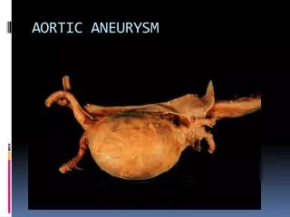
Definition • Aneurysm: irreversible dilation of an artery at least 1.5 times its normal caliber • True aneurysm vs. False aneurysm • Varieties: • Degenerative – due to atherosclerosis, most common type • Traumatic – iatrogenic, catheter-related, penetrating trauma • Poststenotic – Bernoulli’s principle, occurs distally (distal to coarctation, distal to cervical rib in thoracic outlet syndrome, etc.) • Dissecting • Mycotic – infected • Anastomotic – separation between graft and native artery
Abdominal Aortic Aneurysm • Fusiform dilation of abdominal aorta > 1.5 times its normal diameter • Incidence: 5% of elderly population >60 years old (6-9 times more common in males) • Relative risk: 11.6% in patients with first-degree relative with known AAA • Risk factors: Atherosclerosis, HTN, smoking, male gender, advanced age, connective tissue disease • Risk factors for rupture: diastolic HTN, initially large size at diagnosis, COPD, symptomatic, recent rapid expansion
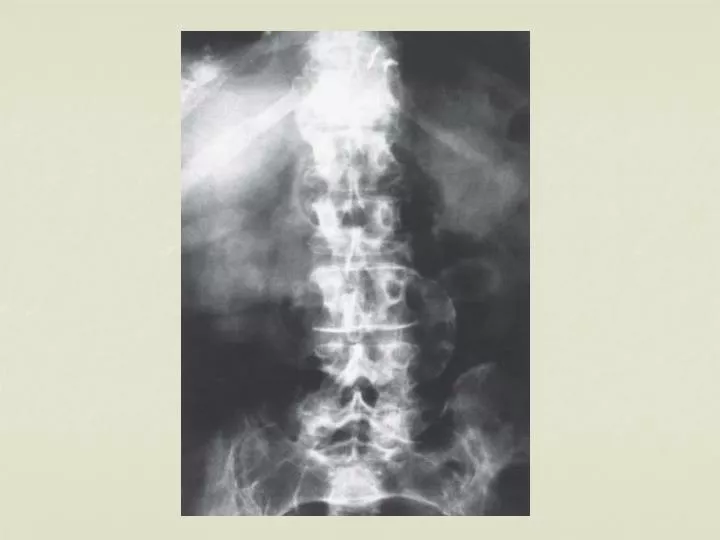
Diagnosis • Exam • Periumbilical palpable pulsatile mass • Ultrasound • Study of choice for initial diagnosis • Used to follow progression of aneurysm over time • Abdominal or back radiographs • Calcifications of aneurysm wall may be seen in ~75% of patients
Diagnosis • CT scan • Character, wall thickness, location with respect to renal arteries, presence of leak or rupture • With Contrast for visualization of surrounding vasculature; essential for planning repair • MRI • Greater detail than CT or US regarding lumen, surface anatomy, neck, relationship to renal arteries • Angiogram • Defines vascular anatomy, assess lumen patency and iliac/renal involvement • Especially important in cases of mesenteric ischemia, HTN, renal dysfunction, horseshoe kidney, claudication
AAA Screening • U.S. Preventive Services Task Force recommends one-time screening by ultrasonography in men age 65 to 75 years who have ever smoked • No recommendation (for or against) screening in men age 65 to 75 who have never smoked, and an explicit recommendation against routine screening in women, based on the relatively low yield • Repeated screening does not appear to be needed
Triad of Rupture • Abdominal pain • Pulsatile abdominal mass • Hypotension