Download
1 / 37
370 likes | 373 Views
This research paper explores the mechanism and regulation of oxidative phosphorylation, the process by which energy release during electron transport is coupled to ATP synthesis. It also discusses the role of uncouplers, the thermogenin protein, and other ways energy can be wasted in this process. Additionally, the paper examines the ATP synthase and its binding change mechanism. Finally, it investigates the regulation of oxidative phosphorylation and the effects of inhibitors in hypoxic conditions.

E N D
BC368 Biochemistry of the Cell II Oxidative Phosphorylation CH 19 (pp 731-768) March 31, 2015
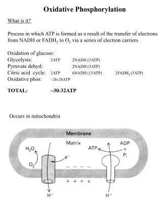
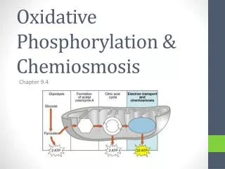
Oxidative phosphorylation is the coupling of energy release during electron transport to ATP synthesis. "Anyone who is not confused about oxidative phosphorylation just doesn't understand the situation." -Efraim Racker 1913-1991
Chemiosmotic Theory Fig 19-19
Proton Motive Force = - Fig 19-17
Case Study In 1933, Stanford biochemists Cutting and Tainter published a report in the Journal of the American Medical Association on the use of dinitrophenol (DNP) to treat obesity. After its first year on the market, an estimated 100,000 people had been treated with DNP in the United States, in addition to many others abroad. Unfortunately, in some cases the treatment eliminated not only the fat, but also the patient. How does DNP work as a diet pill, and what side effects would you expect?
Uncouplers Fig 19-20
Fig 19-34 Uncouplers • Thermogenin dissipates the proton gradient…no work is done. Huffington Post
Fig 19-34 Uncouplers
Other ways to waste energy • Bypassing the proton pumps leads to production of heat instead of ATP
Chemiosmotic Theory Fig 19-19
Mechanism of ATP Synthesis https://www.youtube.com/watch?v=PjdPTY1wHdQ
ATP Synthase: Fo and F1 • In the 1960’s, “lollipop” structures were evident through electron microscopy in samples of everted inner membranes from bovine mitochondria.
ATP Synthase: Fo and F1 Matrix side Matrix side
ATP Synthase: Kinetics Fig 19-24
ATP Synthase: The Binding Change Mechanism • Each β subunit has a different conformation: • β-ADP • β-ATP • β-empty Fig 19-26
ATP Synthase: The Binding Change Mechanism ADP and Pi bind Fig 19-26
ATP Synthase: The Binding Change Mechanism Conformation changes, catalyzing ATP formation; energy provided by H+ movement Fig 19-26
ATP Synthase: The Binding Change Mechanism Conformation changes; ATP dissociates; energy provided by H+ movement Fig 19-26
ATP Synthase: The Binding Change Mechanism Animation: Binding Change Mechanism Conformation changes back to initial state so that cycle continues Fig 19-26
ATP Synthase: The Binding Change Mechanism ATP Synthase
ATP Synthase: Rotation of Fo via the c Ring • Each c subunit has two half-channels, open to either the intermembrane space or to the matrix, that allow protons to access a key Asp residue. • Protonation of the Asp relieves the negative charge and allows rotation into the membrane. • Rotation of negative Asp out of the membrane results in deprotonation. Animation: start at :23
Mitochondrial “shuttles” • Functionally, transport of OH-out is the same as transport of H+in.
Pmf-driven transport Fig 19-30
Malate-Asp shuttle • Liver, kidney, and heart • Results in NADH in the matrix • Complicated, but free! Fig 19-31
Malate-Asp shuttle • Liver, kidney, and heart • Results in NADH in the matrix • Complicated, but free! Fig 19-31
Glycerol 3-P shuttle • Skeletal muscle and brain • Electrons enter at Q. • Easier, but costly!
Regulation Acceptor Control Fig 19-20
Regulation Fig 19-35 Coordinated Control
In-Class Problem The mitochondria of a patient oxidize NADH irrespective of whether ADP is present. The P:O ratio (ATP synthesized per oxygen atom [or pair of electrons]consumed) for oxidative phosphorylation by these mitochondria is less than normal. Predict the likely symptoms of this disorder.
Hypoxia • Normally, the ATP synthase makes ATP, using the proton gradient • Sometimes, the ATP synthase uses ATP to generate a proton gradient (acts as a ATPase). (bacteria or hypoxia) makes
The protein IF1 protects the cell from hypoxia-induced ATP hydrolysis. Hypoxia IF1 inhibitor (a dimer at low pH) Inhibition of ATPase by IF1 Fig 19-33
Hypoxia • When O2 is limiting, electrons may fall out of the electron transport chain, often at Q−.
Hypoxia • When O2 is limiting, electrons may fall out of the electron transport chain, often at Q−. • Superoxide dismutase converts O2− to H2O2. • Glutathione peroxidase breaks down the H2O2.
Hypoxia • Other protective effects are mediated by HIF-1: • Decreased activity of PDH (via the kinase). • Swapping out of a complex IV subunit.
Assign each inhibitor to one of the oxygen traces on the right (the y- axis is [O2]; isolated mitochondria; succinate is the electron source)