Download
1 / 52
520 likes | 654 Views
Gross Anatomy. Background Review. Anatomical Position, terminology . Nomina Anatomica (Latin) Normal anatomical position upright, arms at side, forearm + hand supine . Planes. imaginary lines separating the body at different angles

E N D
Gross Anatomy Background Review
Anatomical Position, terminology • Nomina Anatomica (Latin) • Normal anatomical position • upright, arms at side, forearm + hand supine
Planes • imaginary lines separating the body at different angles • 1. Median - longitudinal - separates left and right (hit surface at anterior, posterior median lines) • 2. Coronal also referred as Frontal - vertical, right angle to median - separate anterior and post (front, back) • 3. Horizontal - (often = transverse, not always) - separate superior & inferior (upper /lower)(transverse of hand is horizontal but the foot is coronal) • 4. Sagittal - vertical off-center, parallel to median (include midsagittal & parasagittal)
Sections / slices • 1. Longitudinal • lengthwise through body or an appendage (direction of its long axis) • can be in median, coronal or sagittal planes • 2. Vertical - same as longitudinal but referring to body in anatomical position • 3. Transverse - cross sections - at right angles to long axis (often horizontal) • 4. Oblique - slanted, at an angle, not in longitudinal or transverse
Relational terms • to localize different structures on the body in anatomical position • 1. Anterior = ventral - toward front (chest, palms, soles); also = rostral in brain • 2. Posterior = dorsal - toward back (dorsum) • 3. Superior = cranial = cephalic - toward head • 4. Inferior = caudal - below, toward feet (tail) • 5. Medial - toward midline or median plane • 6. Lateral - toward side, away from median (little toe = lateral; little finger = *medial) • 7. Intermediate - between 2 structures • 8. visceral • 9. parietal
Relative / Comparison • Terms to describe relative positions of & to compare 2 structures • 1. Proximal - nearer trunk or point of origin • 2. Distal - farther from trunk or point of origin • 3. Superficial - to surface • 4. Deep - away from surface • 5. External - toward exterior, especially of an organ • 6. Internal - to the interior of an organ or inner surface • 7. Ipsilateral - same side of body • 8. Contralateral - opposite side ( Right vs. Left ) • 9. Central - near or towards the center • 10. Peripheral - father, away from the center
Movements - of body parts • 1. Abduction - away from midline • 2. Adduction - toward midline • 3. Flexion - bend - decrease angle of a joint • 4. Extension - straighten - increase angle of a joint (hyperextension = beyond straight point) • 5. Lateral rotation - rotate outward (e.g. leg) • 6. Medial rotation - rotate inward • 7. Circumduction - circular motion (involves 1-4) • 8. Inversion - sole of foot toward median plane
Movements - of body parts • 9. Eversion - sole of foot laterally - outward to side • 10. Pronation - rotate hand so palm faces posterior • 11. Supination - rotate hand so palm is anterior • 12. Protrusion - move anteriorly - stick out (e.g. chin) • 13. Retraction / Retrusion - move posteriorly - pull, tuck in (shoulders, chin) • 14. Elevation - lifting • 15. Depression - lowering to a more inferior position • 16. Opposition - move thumb towards the other digits • 17. Reposition - move thumb away from other digits
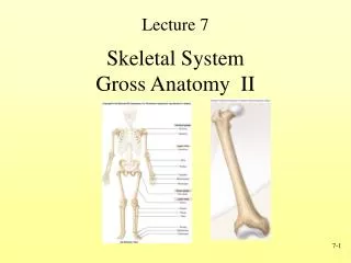
Systems • 1. Integumentary - skin + accessory structures • 2. Skeletal - bones, cartilages • 3. Articular - joints (+ bones, ligaments at each) • 4. Muscular - muscles (part of musculoskeletal) • 5. Nervous - brain, spinal cord, nerves • 6. Circulatory - vascular - heart + arteries, veins, capillaries + lymphatic (nodes, vessels)
Systems • 7. Respiratory - lungs, diaphragm, airways • 8. Digestive - alimentary canal (mouth to anus) + accessory organs (liver, pancreas etc) • 9. Renal / Urinary - kidneys, bladder, excretory tubule system • 10. Endocrine - endocrine glands (pituitary, hypothalamus, adrenals, reproductive glands) • 11. Reproductive - ovaries or testes (a.k.a. urogenital system, esp. in males)
Muscular System • function in movement, support (posture), heat generation
Skeletal Muscle • 1. striated, voluntary • 2. moves bones, skin (facial muscles) - typically, origin & insertion are attached across a joint • - most are under our control, although many movements are reflexes - eg stretch reflex • 3. attachments: • each has an Origin = proximal attachment & Insertion = distal attachment • 4. structure: a. muscle fiber = muscle cell = structural unit b. motor unit = one motor neuron + all muscle fibers under its control • size varies inversely with precision, delicacy of control
Skeletal muscle • 5. movements: a. agonists = prime movers - carry out the main movement • b. antagonists - oppose action of agonists - relax as agonist contracts for smooth movement • c. synergists - complement/ work with/ support prime movers - especially to support the joint • d. fixators - steady proximal part of limb while distal part is moved (e.g. forearm vs. hand)
Smooth muscle • 1. non-striated, involuntary - controlled by ANS • 2. propels foodstuffs thru alimentary canal; blood thru vessels a. undergoes peristalsis - rhythmic waves of contraction • b. maintains a constant level of tone (esp. important in vasculature)
Cardiac muscle • 1. striated, but involuntary - spontaneous excitation; under control of ANS • 2. pumps blood through heart
Nervous System • Major Functions of the Nervous System • 1. Sensory - many receptor types in body sense, detect changes in body or surroundings • 2. Integration - of information received thru sensory system to arrive at a proper response • 3. Motor - nerve impulses trigger responses by the body’s effectors = muscles, glands etc
Nervous System • Divisions • 1. CNS = the brain & spinal cord - nerves do NOT regenerate after injury • 2. PNS = peripheral nerves that communicate between the CNS and the rest of the body- nerves MAY regenerate after injury
Structures • 1. Neurons = the functional cells of the NS - transmit electrical impulsesa. dendrites = receptive processes; receive impulses from receptors & other neuronsb. axon = single transmitting process, sends impulse to other neurons or to effectors- myelin sheath - insulates axon - speeds transmission- nodes of Ranvier - spaces between sections of myelin
Structures • 2. Neuroglial cells • a. supportive accessory cells - insulate, connect neurons, attach neurons with surrounding tissue • b. provide nutritive support - to provide energy, provide central nervous system with blood • 3. Types of Neurons - Classified by Function: • a. Motor • b. Sensory • c. Interneurons depends on how information flows
Basic terminology • 1. Nucleus - a group of neuronal cell bodies within the CNS • 2. Ganglion (ganglia, pl) - group of cell bodies outside the CNS • 3. Nerve - collection of axons (fibers) in PNS ; fasciculus - a bundle of nerves- plexus = network of nerves in one area • 4. Tract - bundle of axons/fibers in the CNS • 5. Gray matter - concentrations of cell bodies in CNS, e.g.. cerebral cortex • 6. White matter - axons, processes in the CNS
Connective tissue meninges • 1. CNS = meninges: • connective tissue surround, protect the nervous sytem • a. pia mater - immediately next to the nervous tissue • b. arachnoid - middle layer • the 2 inner layers, leptomeninges, thin & delicate • c. dura mater - outermost meninges, thicker & very tough • d. cerebrospinal fluid fills space between arachnoid and pia mater
PNS • a. endoneurium: thin collagenous layer, immediately surrounds a myelinated n fiber • b. perineurium: CT covering surrounding a fascicle of nerve fibers • c. epineurium: thick CT layer surrounding many fascicles wh make up a nerve trunk • * the 3 CNS meningial layers are continuous with the CT layers around PNS nerves
The Peripheral Nervous System • 1. Cranial nerves: 12 pair transmit from brain to head, neck, trunk (mixed, motor & sensory, more in Neuro) • 2. Spinal nerves: 31 pair communicate btw spinal cord & neck, trunk, arms, legs (mixed) • a. Dorsal roots - sensory/afferent fibers into cord, dorsal root ganglia contain cell bodies of sensory neurons • b. Ventral roots - motor/efferent ff from cord to periphery, branch once outside cord • c. dorsal & ventral roots combine, form spinal nerve, which branches again: • dorsal primary rami: supply fibers to dorsum (back) • ventral primary rami: supply fibers to anterolateral trunk, limbs
Somatic Nervous System • 1. nerves that communicate w skin & skeletal muscles • 2. functions are under conscious control
Autonomic Nervous System • 1. concerned with automatic/ visceral functions, homeostatic mechanisms (CVS, digestion) • 2. function without conscious control • 3. control the function of visceral organs - heart, smooth muscle, vessels, glands D. ANS has 2 Divisions
Sympathetic Division • concerned primarily w survival, emergency, stressful situations – “Fight or flight system” • a. connects with thoracic & lumbar segments: T1 - L2 or 3 • b. many connect with Peripheral NS efferent/motor neuron in ganglia: • paravertebral ganglia (sympathetic trunk) • prevertebral ganglia (visceral ganglia., e.g. celiac ganglia.) • the adrenal medulla (exception, innervated by preganglionic fiber directly) • c. route: spinal cord lateral horn - sympathetic axons (preganglionic) => through ventral root => through white ramus communicante (branch to ganglion) => to paravertebral ganglia
Sympathetic Division • then either: 1) passes through ganglia (without synapse) directly to viscera (as a splanchnic nerve) • 2) ascends or descends through trunk to another level, then postganglionic fibers innervate organs like heart, lungs, glands • 3) synapses in ganglia with excitor neurons => postganglionic go back through gray ramus communicante => then blend in with spinal nerve > effectors
Parasympathetic system “Breed and Feed” a. concerned w normal functions: eating, sleeping, procreation, conserving energy b. connects with cranial & sacral segments: cranial = III, VII, IX, X & spinal = S2 - S4
Spinal cord • 1. continuation of the CNS from the brain, out of the skull into the vertebral column • 2. composed of 31 segments - a pair of spinal nerve exits each segment, to the periphery • 3. gray matter localized to central core: mostly cell bodies, proximal unmyelinated process’ • 4. white matter surrounds central area: composed of primarily myelinated axons, fibers
Spinal cord • 5. Function - spinal cord communicates to & from the brain & the remainder of the body • a. ascending tracts carry sensory input to CNS (some to spinal cord, some to brain) • b. descending tracts leaving the brain & spinal cord carry motor output to effectors • c. most fiber tracts cross to contralateral side at some point in spinal cord or brain
mixed nerves • 31 pairs • 8 cervical (C1 to C8) • 12 thoracic (T1 to T12) • 5 lumbar (L1 to L5) • 5 sacral (S1 to S5) • 1 coccygeal (Co)