Download

1 / 28
280 likes | 387 Views
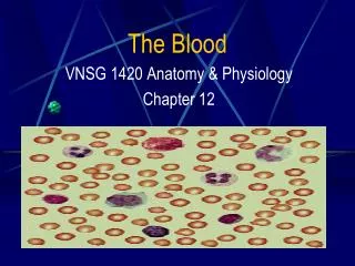
The Blood. The Blood. Definition Blood is a connective tissue, made of fluid (plasma) and cellular elements (RBC, WBC, and platelets) Its volume is 5-6 L in males and 4-5 L in females It is slightly alkaline, with a pH of ~ 7.4

E N D
The Blood Definition Blood is a connective tissue, made of fluid (plasma) and cellular elements (RBC, WBC, and platelets) Its volume is 5-6 L in males and 4-5 L in females It is slightly alkaline, with a pH of ~ 7.4 Its color varies from bright to dark red It has a salty metallic taste
The Blood Functions The bloodis an organ that reaches all the other tissues Transports oxygen and nutrients Removes CO2 and other by-products of cell activity Participates in the defense against infection Participates in hemostasis Participates in body heat distribution and regulation
Marieb and Hoehn Human Anatomy & Physiology seventh edition Pearson Benjamin Cummings
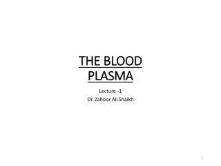
The Blood Plasma Straw colored fluid made of water (~90%), other contents include: Proteins make the bulk of the solutes: Albumens (60%), manufactured in the liver are the most abundant Globulins (36%) are immune bodies Fibrinogen (4%) for blood clotting Nutrients: glucose, amino acids, lipids, cholesterol Electrolytes: Na+, K+, Ca++, Mg++, H+, Cl-, HCO3-, PO4--, SO4-- Waste: urea, creatinine, uric acid, bilirubin Gases: O2 , CO2 , N2 Protein Plasma without clotting factors is called “serum”
Peripheral blood smear Marieb and Hoehn Human Anatomy & Physiology seventh edition Pearson Benjamin Cummings
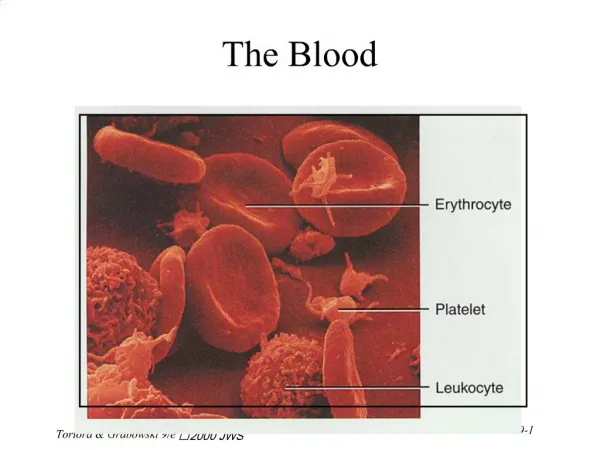
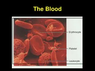
Electron micrograph of blood smear Pathophysiology McCance & Huether fifth edition Elsevier Mosby
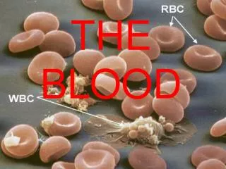
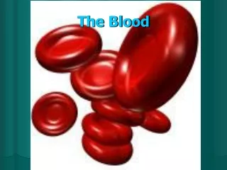
The Blood RBC An RBC is a 7.5 micron disc shaped body with a central depression NO nucleus NO mitochondria AN RBC contains hemoglobin Antigens on RBC cell membrane determine blood type RBCs function is gas exchange: O2 to the tissues and CO2 to the lungs
The Blood RBC Development of RBCs Hypoxia → erythropoietin (kidney)→ red marrow of long bones→ The normal number of RBCs is 4.3-5 million/mm3 in the female and 5.1-5.8 million/mm3 in the male The normal values for Hgb are 13-15 gm/dl for females and 14-16 gm/dl in males Amino acids, lipids, carbohdrates, iron, vitamin B12 and folic acid are essntial for hemoglobin synthesis
The Blood RBC Hemoglobin has a remarkable ability to bind with oxygen forming oxyhemoglobin It can also release the oxygen to the tissues becoming deoxyhemoglobin
The Blood RBC Destruction Life span ~ 120 days RBCs are phagocytosed by the reticulo-endothlial cells of the spleen
The Blood RBC Disorders of RBCs Anemia is reduced RBC count Anemias can be caused by RBC loss or reduced production Hemorrhage, hemolysis, depressed bone marrow Reduced hemoglobin content of RBCs Iron, intrinsic factor, folic acid, or B12 deficiency Congenital hemoglobin defects Thalassemia, sickle cell anemia, spherocytosis
Normal and sickle cell RBC In sickle cell disease hemoglobin S replaces the β chain In thalassemias, the α or β chains can be absent or defective Marieb and Hoehn Human Anatomy & Physiology seventh edition Pearson Benjamin Cummings
The Blood White Blood Cells (WBC) Function Lymphocytes are the effectors of the immune function WBCs main function is to fight bacterial infections WBCs are the only nucleated blood formed elements They exercise their functions in the tissues not the blood stream WBC migrate to the tissue spaces WBC destroy the bacterial cell wall Life span 1 – 9 days
The Blood White Blood Cells (WBC) Lymphocytes, the smallest and second most abundant T cells (80%) mediate cellular immunity B cells mediate humoral immunity Monocytes, the largest, migrate to the tissues and become macrophages involved in cellular immunity
White blood cells, the granulcytes Davidson’s The Priciples and Practice of Medicine, eigthteenth edition Churchill Livingstone
Monocytes and lymphocytes Davidson’s The Priciples and Practice of Medicine, eighteenth edition Churchill Livingstone
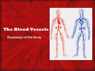
The Blood Blood Coagulation Coagulation is a natural mechanism that acts to diminish blood loss from hemorrhage Coagulation occurs in three Stages Platelet plug The cascade leading to fibrin (clot) formation Clot retraction and repair (PDGF)
The Blood Blood Coagulation The Platelets Platelets are cell fragments Their life span is 8-14 days
The structure of a platelet Davidson’s The Priciples and Practice of Medicine, eighteenth edition Churchill Livingstone
Initial vasoconstriction and platelet plug v Vander’s Physiology eighth edition Mc Graw Hill
Intrinsic and extrinsic coagulation pathways Calcium ions are essential for the coagulation cascade Vander’s Physiology eighth edition Mc Graw Hill
The role of the liver in blood coagulation Vander’s Physiology eighth edition Mc graw Hill
EM of a blood clot: RBC’s and fibrin NIBSC?Science Photo Libraray – Taken from Vander Physiology eighth edition Mc Graw Hill
Blood Types RBC’s have surface antigens RBC’s can be grouped according to the presence or absence of certain antigens There are many RBC antigens but only a A, B, AB and the Rh are of clinical significance Each one of the four types can be Rh positive or negative Donor blood is mixed with recipient serum to decide compatibility, donor cell clumping indicate incompatibility The Blood
Major blood groups A certain group posseses its antigen on the RBC surface and antibodies against the others Marieb and Hoehn Human Anatomy & Physiology seventh edition Pearson Benjamin Cummings
The Blood Rh Factor There are no preformed Rh antibodies in Rh- individuals They develop after exposure to Rh+ factors (antigens) This explains the fact that an Rh+ fetus of an Rh- mother does not usually suffer But the Rh- mother can be sensitized to the Rh+ antigens during the first pregnancy, especially during delivery Subsequent fetuses can suffer from the mother’s Rh antibodies passed to it and its blood hemolyzes This can be prevented by giving the mother serum that blocks the Rh+ factors antigenicity
The Blood Rh Factor Eighty five percent of the population have Rh antigens (Rh+) There are several Rh groups (factors, antigens) Three groups: Rh, C, D, and E, are of clinical importance An Rh- mother may develop antibodies against her Rh+ fetus The Rh- mother antibodies can pass to the Rh+ fetus resulting in hemolysis of its RBCs, a condition know as erythroblastosis fetalis This sequence usually occurs after the first pregnancy