Download
1 / 14
140 likes | 217 Views
Legend to Supplementary Figure 1. Histograms showing the number of times that each amino acid undergoes: (a) synonymous (b) nonsense and (c) missense substitutions. The two series in (c) show the

E N D
Legend to Supplementary Figure 1. Histograms showing the number of times that each amino acid undergoes: (a) synonymous (b) nonsense and (c) missense substitutions. The two series in (c) show the number of times that each amino acid: (i) undergoes and (ii) is generated as a result of missense mutations. In each of the five histograms, (d) to (h), missense mutations undergone by 4 amino acids (black bars) have been examined in detail. In each figure, each black bar shows the total number of times that a WT amino acid undergoes missense mutations; the striped bars following the black bar show the frequencies of the mutant amino acids replacing the WT amino acid as a result of missense mutations.
Supplementary Figure 1 (a) (b) R, Q, E G, L G (c) G C,K,N C K N
PS,L P L S AT,V YC (d) (e) A Y T V C Supplementary Figure 1 (f) (g) (h) EK GR E G K R
Supplementary Figure 2 Legend to Supplementary Figure 2. Distribution of WT and mutant pairs of (adjacent) bases in 2-base substitutions. The frequency of occurrence of each WT and mutant pair is shown, respectively, in the first and second series.
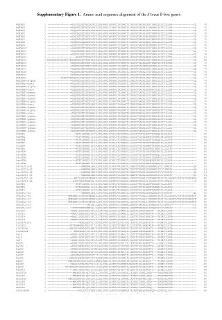
Legend to Supplementary Figure 3. (A) The different possible ways in which nt can be deleted in I-F and FS deletions. The column headings indicate whether base(s) in a single codon or in multiple codons are being deleted. 1, 2, 3 in the row and column headings refer to codon positions. The row heading indicates the type of deletion. For each type of deletion, there are 2 lines. The first line gives the WT codon(s). In the second line, deleted base(s) are represented as ‘–‘, and start and end of deletion are each marked by ‘|’. (a) In I-F deletions, nt are deleted in multiples of 3 (3, 6, 9, 12…) and, therefore, no change in the gene reading frame occurs. The deletions are of the types, 1-3, 2-1, 3-2. 1-3 type ones are complete codon deletions that begin at pos1 and end at pos3 of the same or a downstream codon. 2-1 type ones begin at pos2 of one codon and end at pos1 of the adjacent or a downstream codon. (b) In FS deletions, odd or even numbers of nt, that are not multiples of 3 (1, 2, 4, 5, 7…), are deleted and, as a result, the gene reading frame changes. The deletions are of 6 types: 1-1, 2-2, 3-3, 1-2, 2-3, 3-1. In a 1-1 type one, either a single nt at pos1 of a single codon is deleted or the deletion begins at pos1 of one codon and ends at pos1 of the next or a more downstream codon. In a 1-2 type one, either 2 adjacent nt, occurring at pos1 and pos2 of a single codon, are deleted or the deletion begins at pos1 of one codon and ends at pos2 of the next or a more downstream codon. 3-1 type deletions are always multiple codon deletions. (B) The different possible ways in which nt can be inserted in I-F and FS insertions. The column headings indicate whether base(s) are being inserted in a single codon or in multiple codons. 1, 2, 3 in the row and column headings refer to codon positions. The row heading indicates the type of insertion. For each type of insertion, there are 2 lines. The first line gives the WT codon(s). In the second line, the start and end of insertion are each marked by ‘|’, the inserted base(s) are shown underlined, and the WT bases are given on either side of the insertion. (a) I-F insertions cause no change in the gene reading frame because nt are inserted in multiples of 3. They are of the types: 1-3, 2-1, 3-2. In 1-3 type ones, the inserted nt constitute complete codon(s). In 2-1 type ones, the inserted nt constitute the segment running from pos2 of one codon to pos1 of the adjacent or a more downstream codon. (b) FS insertions cause a change in the gene reading frame because odd or even numbers of nt, that are not multiples of 3, are inserted. They are of the types: 1-1, 2-2, 3-3, 1-2, 2-3, 3-1. In a 1-1 type insertion, either a single inserted nt constitutes pos1 of a codon or the insertion constitutes the segment running from pos1 of a codon to pos1 of a downstream codon. In a 1-2 type one, either 2 inserted nt constitute pos1 and pos2 of a single codon, or the insertion constitutes the segment running from pos1 of a codon to pos2 of a downstream codon. In a 3-1 type one, the inserted nt always constitute parts of more than one codon.
In-frame deletions - no change in gene reading frame; Thus nt have to be deleted in multiples of 3 (3, 6, 9, 12, ...); nt in single codon nt in multiple codons 1 2 3 1 2 3 1 2 3 1-3 t c t t c t a t t │- - -││- - - - - -│ 2-1 t c a c g a t│- - -│g a 3-2 c t t c a g c t│- - -│g Frameshift deletions - change in reading frame; odd or even nos. of nt, that are NOT MULTIPLES OF 3, are deleted (1, 2, 4, 5, 7,...) nt in single codon nt in multiple codons 1 2 3 1 2 3 1 2 3 1-1 g a c g a c a t t - a c │- - - -│t t 2-2 c c t c c t c g a c - t c│- - - -│a 3-3 a g t a g t c a g a g - a g│- - - -│ 1-2 c t a c t a t c t │- -│a │- - - - -│t 2-3 t t t t t t t c a t - - t│- - - - -│ 3-1 g a t a g t g a│- -│g t Supplementary Figure 3A (a) (b)
In-frame insertions - no change in gene reading frame; Thus nt have to be inserted in multiples of 3 (3, 6, 9, 12, ...); nt in single codon nt in multiple codons 1 2 3 1 2 3 1 2 3 1-3 t c t t c t a t t │cgg│ t c t│ctgtcg│ t c t a t t 2-1 t c a c g a t│gga│c a c g a 3-2 c t t c a g c t│gag│t c a g Frameshift insertions - change in reading frame; odd or even nos. of nt, that are NOT MULTIPLES OF 3, are inserted (1, 2, 4, 5, 7,...) nt in single codon nt in multiple codons 1 2 3 1 2 3 1 2 3 1-1 g a c g a c a t t │a│g a c │ggtg│g a c a t t 2-2 c c t c c t c g a c│a│t c│agga│c t c g a 3-3 a g t a g t c a g a g│g│ a g│ttat│ t c a g 1-2 c t a c t a t c t │ac│a │atgtc│c t a t c t 2-3 t t t t t t t c a t│at│ t│agtga│ t t t c a 3-1 g a t a g t g a│ta│t a g t Supplementary Figure 3B (a) (b)
Supplementary Figure 4 Legend to Supplementary Figure 4.Histograms showing distributions of the locations of: (A) deletions and (B) insertions in proteins. In each histogram, the two series show the distributions for I-F and FS deletions or insertions. The X-axis indicates the locations of deletions or insertions, and the Y-axis, the frequency of occurrence of deletions or insertions at each location. In B, the last 2 sets of bars give the frequencies with which proteins become shorter or longer as a result of FS and I-F insertions. (First_1/3, N-terminal region; second_1/3, middle region; third_1/3, C-terminal region; bet_1_2, between first and second regions; bet_2_3, between second and third regions; mid_gone, middle region deleted; prot_gone, entire protein deleted; no_n_ter, N-terminal residues deleted; no_c_ter, C-terminal residues deleted). A B
Supplementary Figure 5(A-B) Legend to Supplementary Figure 5. Distributions of lengths of protein lost or gained due to: (A) deletions and (B) insertions. In each histogram, the two series show the distributions for FS and I-F deletions or insertions. Length of protein lost/gained due to each indel was calculated as: (length of WT protein - length of mutant protein). In (A), lengths of protein lost are given as intervals along the x-axis; the intervals are not all of equal length. In (B), negative and positive values, along the x-axis, indicate lengths of protein lost and gained, respectively. The observed frequency with which lengths of protein are lost or gained in each interval is given along the y-axis. (A) (B) increase in prot length decrease in prot length
Supplementary Figure 6 Legend to Supplementary Figure 6. Length distribution of frame-shifted or corrupted sequences resulting from FS deletions and FS insertions (first and second series, respectively). Frame-shifted sequence lengths in amino acids are given as intervals along the x-axis, and the number of occurrences of frame-shifted sequences in each interval is given along the y-axis.
Legend to Supplementary Figure 7. Histograms showing, for each mutation type, the distribution of mutation positions over the lengths of proteins. While the distribution for all mutation positions considered together is shown in Figure 8, those for individual types of mutations are shown here. Distributions are shown in (a, b, c) for missense, nonsense and synonymous substitutions, in (d, e) for I-F and FS deletions, and in (f,g) for I-F and FS insertions. For a key to the histograms, see legend to Figure 8. As an illustrative example, in histogram (a), in the PO, JAK2, 85% of all missense mutation positions (second bar) occur in 18% of the protein length (first bar).
Supplementary Figure 7 (a) (c) (b)
Supplementary Figure 7 (d) (e)
Supplementary Figure 7 (f) (g)