Download
1 / 46
510 likes | 1.46k Views
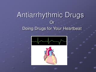
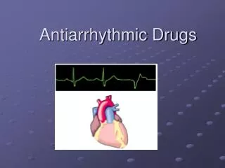
Antiarrhythmic drugs . Conducting system of the heart. Sequence of cardiac excitation. Figure 12-11. The Bundle of His and other parts of the conducting system deliver the excitation to the apex of the heart so that ventricular contraction occurs in an upward sweep. .

E N D
Sequence of cardiac excitation Figure 12-11 The Bundle of His and other parts of the conducting system deliver the excitation to the apex of the heart so that ventricular contraction occurs in an upward sweep. The sinoatrial node is the heart’s pacemaker because it initiates each wave of excitation with atrial contraction.
Normal Sinus Rhythm • Heart rhythm is determined by SA node = Cardiac Pacemaker • Called sinus rhythm • Specialised pacemaker cells spontaneously generate APs • APs spread through the conducting pathways • Normal sinus rate 60-100 beats/min
Conducting System • SA Node AP triggers atrial depolarisation • AV Nnode- Only pathway for AP to enter ventricles • Conducts slowly: Complete atrial systole before ventricular systole • Conducts rapidly through His Bundles & Purkinje – Ventricular depolarization & contraction
Conducting System • Permits rapid organized depolarization of ventricular myocytes • Necessary for the efficient generation of pressure during systole • Atrial activation complete 0.09s after SAN firing • Delay at AVN • Septum activated 0.16s • Whole ventricle activated by 0.23s
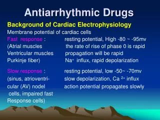
Cardiac Action Potential • Phase 4: resting membrane potential • AP depolarizes cells to threshold -70mV • Phase 0: Rapid depolarization • Caused by a transient opening of fast Na channels • Increases inward directed depolarizing Na+ currents • Generate "fast-response" APs
Cardiac Action Potential • Phase 1: Initial repolarization • Open K channel: transient outward hyperpolarizing K+ current • Large increase in slow inward Ca++ occurs at the same time • L-type Ca Ch open -40mV • Repolarization delayed • Phase 2: Plateau phase • Plateau phase prolongs AP duration vs APs in nerves and skeletal muscle
Cardiac Action Potential • Phase 3: Repolarization • K channels open • Inactivation of Ca++ channels • Action potential in non-pacemaker cells is primarily determined by relative changes in fast Na+, slow Ca++ and K+ conductances and currents
Refractory Periods • Once an AP is initiated, there is a period (phase 0,1,2, part 3) that a new AP cannot be initiated. • Effective or Absolute refractory period (ERP or ARP) • Stimulation of cell by adjacent cell depolarizing does not produce new propagated APs • Prevents compounded APs from occurring & limits frequency of depolarization and HR
SAN Pacemaker Potential • Fully repolarized -60mv • No stable Resting Membrane Potential • Phase 4: Spontaneous depolarization or pacemaker potential • Slow, inward Na+ channels open - "funny" currents • Cause the membrane potential to begin to spontaneously depolarize • During Ph4 there is also a slow decline in the outward movement of K+
SAN Pacemaker Potential • -50mV T-type CaCh open • Ca in: further depolarizes • -40 mV L-type CaCh open • More Ca in: further depol • AP threshold -35mV • Phase 0: Depolarization • Primarily caused by Ca++ conductance through the L-type Ca++ channels • Movement of Ca++ through these is slow so the rate of depolarization (Phase 0 slope) is slower than in other cardiac cells
SAN Pacemaker Potential • Phase 3: Repolarization • K+ channels open • Increase the outward hyperpolarizing K+ currents • At the same time the L-type Ca++ channels close • gCa++ decreases • Inward depolarizing Ca++ currents diminish • Repolarization
Regulation of Cardiac APs • SNS - Increased with concurrent inhibition vagal tone: • NA binds to B1 Rec • Increases cAMP • Increases Ca and Na in • Decreases K out • Increases slope phase 0 • Non-Nodal tissue: • More rapid depolarisation • More forceful contraction • Pacemaker current (If) enhanced • Increase slope phase 4 • Pacemaker potential more rapidly reaches threshold • Rate increased
Regulation of Cardiac APs • PSNS (Vagal N) • Ach binds M2 rec • Increases gK+ • Decreases inward Ca & Na • Non-Nodal tissue: • More rapid depolarisation • More forceful contraction • Pacemaker current (If) suppressed • Decreases pacemaker rate • Decrease slope of Phase 4 • Hyperpolarizes in Phase 4 • Longer time to reach threshold voltage
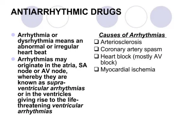
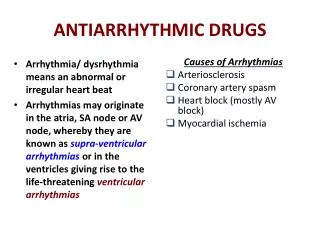
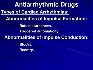
What is an Arrhythmia ? • Irregular rhythm • Abnormal Rate • Conduction abnormality
What causes an arrhythmia? • Changes in automaticity of the Pace Maker • Ectopic foci causing abnormal Aps • Excitable group of cells that causes a premature heart beat outside the normally functioning SA node of the human heart • Hypoxic/Ischaemic tissue can undergo spontaneous depolarisation and become an ectopic pacemaker • Reentry tachycardias • Block of conduction pathways • Abnormal conduction pathways (WPW) • Electrolyte disturbances and DRUGS
Re-Entry Mechanism • Branch 2 has a unidirectional block • Impulses can travel retrograde (3 to 2) but not orthograde. • An AP will travel down the branch 1, into the common distal path (br 3), then travel retrograde through the unidirectional block in branch 2. • When the AP exits the block, if it finds the tissue excitable, it will continue by traveling down (reenter) the branch 1. • If it finds the tissue unexcitable (ERP) the AP will die. • Timing is critical –AP exiting the block must find excitable tissue to propagate. • If it can re-excite the tissue, a circular pathway of high frequency impulses (tachyarrhythmia) will become the source of APs that spread throughout a region of the heart (ventricle) or the entire heart.
Notes • Anti-arrhythmics are also pro-arrhythmics Dangerous side effects
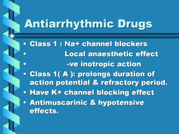
Class I =sodium channel blockers • Interfere with the sodium channel • Grouped by what effect they have on the Na+ channel, and what effect they have on cardiac action potentials • 1A lengthens the action potential (right shift) • 1B shortens the action potential (left shift) • 1C does not significantly affect the action potential (no shift) • Called Membrane Stabilizing agents. • The 'stabilizing' is the word used to describe the decrease of excitogenicity of the plasma membrane which is brought about by these agents
Sodium channels physiology • Deactivated (closed) • Activated (open) • inactivated (closed) .
Class I A Agents • Block open ACTIVATED Na channels • Greater affinity for rapidly firing channels • Slow phase 0 depolarisation - upstroke of AP • Lengthen APD and ERP. • Cause torsades de pointes • Also blocks K Ch • Prolong QRS duration on ECG • Uses: • Ventricular and supraventricular tachycardia • AnticholinergicS/E • They can cause AV block
Class Ia agents • Disopyramide: • Negative Inotrope • Worsening of heart failure • Strong anticholinergic SE • Quinidine: • Also antimalarial • Procainamide : • Cause systemic lupus erythromatous
Class I B Agents • Block INACTIVATED Na channels • Slow phase 0 depolarisation- Slows upstroke of AP • Shorten APD and ERP • Ratio ERP/APD is increased • Greater affinity for ischaemic tissue that has more inactivated channels, little effect on normal cells – dissociates quickly (0.5sec)
Class Ib agents • Lignocaine(lidocaine): • Also local anasthetic • Used Ventricular arrhythemias in MI pts • Not the best now we use amiodarone • IV Administration • Side effects • CNS SE:convulsions,slurred speech • Induce arrhythmia • Phenytoin • Also anticonvulsant • Drug of choice in treatment of digoxin toxicity • Side effects: • CNS SE:convulsions,slurredspeech,nystagmus • Gingival hyperplasia
Class I C Agents • Block Na channels. • Most potent Na channel block • Dissociate very slowly (10-20 sec) • Strongly depress conduction in myocardium • Slow phase 0 depolarisation - upstroke of AP • No effect on APD • No effect on QRS
Class Ic agents • Flecainide: • Used for life threatening ventricular arrhythemia • Last line treatment • Side effects • Induce arrhythmia • Propafenone • Used for ventricular arrhythmia and supraventricular arrhythmia • Has beta blockage activity • Side effects • Induce arrhythmia • Beta blockage side effects
Class II Agents • Beta Blockers - Block B1 receptors in the heart • Decrease Sympathetic activity • Non-Nodal Tissue: • Increase APD and ERP • SA and AVN: • Decrease heart rate • Decrease conduction velocity (Block re-entry) • Inhibit aberrant PM activity
Class II agents • Propranolol • Atenolol • Esmolol • Metoprolol • Use: • Prevent arrhythmias after MI • Used for supraventricular and ventricular arrhythmia • Esmolol is used for acute surgical arrhythemias
ATENOLOL • Non-selective B-Blocker (B1 and B2) • Indications: Convert or Slow rate in SVTs • 2nd line after Adenosine/Digoxin/Diltiazem • IV atenolol 5 mg over 5 minutes • Repeat to maximum 15 mg. • 50 mg PO BID if IV works • Contraindiactions: • Asthma • CCF. Poor EF. High degree heart block. • Ca channel blockers. Cocaine use.
Class III Agents • Anti-Fibrillatory agents. • Block K channels • Prolong repolarisation • Prolong APD and ERP • Useful in Re-Entry tachycardias
Class III agents • Sotalol • Amiodarone • Uses: • supraventricular and ventricular arrhythmia
Amiodarone • Related structurally to thyroid hormone • Exhibits properties of class I to IV but it is predominantly type III • Taken PO or IV • Side effects: • Photosensitivity • Thyroid disorders • Pulmonary alveolitis • Neuropathy • Blue skin discoloration caused by iodine • Corneal microdeposits • Hepatocellular necrosis
Class IV Agents • Calcium Channel Blockers • Bind to L-type Ca channels in Vascular Smooth Muscle, Cardiac nodal & non-nodal cells • Decrease firing rate of aberrant PM sites • Decrease conduction velocity • Prolong repolarisation
VERAPAMIL • Narrow complex tachycardias • Terminates PSVT/SVT • Rate control in AFib/Aflutter • NOT WPW or VT or high degree block • NOT with BBlockers • Negative Inotropy • Vasodilation – Hypotension • Diltiazemless adverse effects
What does Adenosine Do? • Purine nucleoside • Acts on A1 adenosine receptors • Opens Ach sensitive K channels • Inhibits Ca in current – Suppresses Ca dependent AP (Nodal) • Increases K out current – Hyperpolarisation • Inhibits AVN > SAN • Increases AVN refractory period
ADENOSINE • Interrupts re-entry and aberrant pathways through AVN – Diagnosis and Treament • Drug for narrow complex PSVT • SVT reliant on AV node pathway • NOT atrial flutter or fibrillation or VT • Contraindications: • VT – Hypotension and deterioration • High degree AV block • Poison or drug induced tachycardia • Bronchospasm but short DOA
What does Digoxin Do? • Cardiac glycoside • Blocks Na/K ATPase pump in heart • Less ECF Na for Na/Ca pump • Increased IC Ca • Inotropic: Increases force of contraction • AVN increased refractoriness • Decreases conduction through AVN and SAN • Negative chronotrope: Slows HR Reduces ventricular response to SVTs
Magnesium • Mechanism of action is unknown • Used for treatment of torsades de pointes • Side effects: • Bradycardia • Respiratory paralysis • Flushing • Headache