Download
1 / 32
380 likes | 439 Views
Clinical Pharmacology of Antiarrhythmic Drugs. ANTIARRHYTMIC DRUGS.

E N D
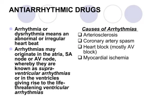
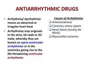
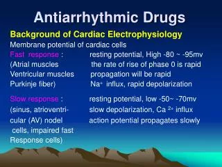
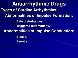
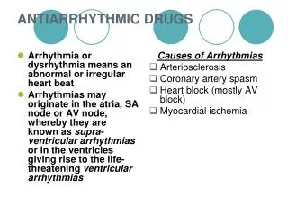
ANTIARRHYTMIC DRUGS Antiarrhythmic agents are a group of pharmaceuticals that are used to suppress abnormal rhythms of the heart (cardiac arrhythmias), such as atrial fibrillation, atrial flutter, ventricular tachycardia, and ventricular fibrillation. These drugs were classified into various groups by SINGH VAUGHAN WILLIAMS as follows:
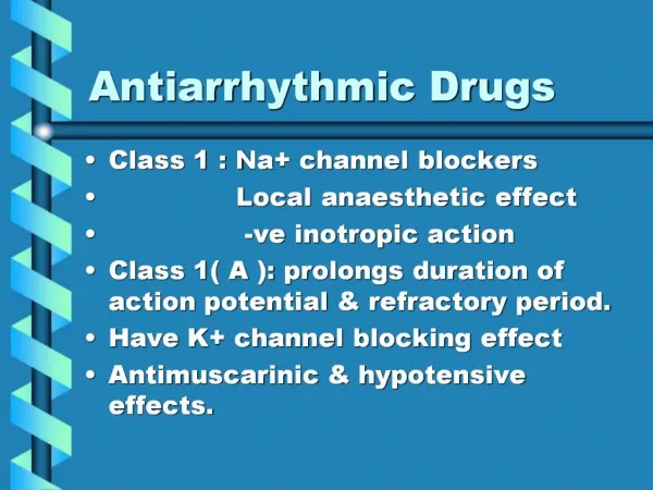
Classification of Anti-arrhythmic Drugs • Na+ channel blockers (membrane stabilizing group). • Beta- adrenergic blockers • K+ channel blockers (prolong repolarisation and action potential) • Ca2+ channel blockers L-type
Class I drugs are divided into three subgroups: Subgroup I A Subgroup I B Subgroup I C
Class I A Antiarrhythmic drugs Also known as fast channel blockers affect QRS complex. Examples: Quinidine, procainamide
Class I B Anti arrhythmic drugs These subgroup of antiarrhyhtmic drugs do not affect QRS complex. Examples: Lidocaine, phenytoin
Class I C Anti arrhythmic drugs Examples: Propafenone, moricizine Mechanism of action: sodium channel blockers Clinical uses: Prevents paroxysmal atrial fibrillation, treats of recurrent tachyarrhythmias of abnormal conduction, contraindicated after myocardial infarction.
Class II anti arrhyhtmic drugs( Beta adrenergic receptors blocker) Mechanism of action: Decrease adrenergic influence on heart by blocking Beta-adrenoreceptors. Clinical uses: decrease myocardial infarction mortality, treatment of supraventricular and vetricular tachyarrhythmia Example: Anaprillinum, metoprolol, atenolol, nadolol, timolol
Class III antiarrhythmic drugs(Potassium channel blocker) Mechanism of action: It prolongs action potential duration and refractory period due to block of potassium channels. Clinical uses: used for treatment of atrial flutter,Wolf-Parkinson-White syndrome, atrial fibrilllation, ventricular tachycardia. Example: Amiodarone, sotalol, bretyllium
Class IV anti arrhythmic drugs(Calcium channel blockers) Mechanism of action: blocks calcium channels decrease conduction through AV node and shorten action potential. Clinical Uses: Hypertension, angina, atrial fibrillation, paroxysmal tachycardia. Examples: Verapamil, dilthiazem
Sinus tachycardia (also colloquially known as sinus tach or sinus tachy) is a heart rhythm with elevated rate of impulses originating from the sinoatrial node, defined as a rate greater than 100 beats/min (bpm) in an average adult.
DIAGNOSIS Usually apparent on the EKG, but if heart rate is above 140 bpm the P wave may be difficult to distinguish from the previous T wave and one may confuse it with a paroxysmal supraventricular tachycardia or atrial flutter with a 2:1 block. Ways to distinguish the three are: Vagal maneuvers (such as carotid sinus massage or Valsalva's maneuver) to slow the rate and identification of P waves administer AV blockers (e.g., adenosine, verapamil) to identify atrial flutter with 2:1 block
TREATMENT Not required for physiologic sinus tachycardia. Underlying causes are treated if present. Acute myocardial infarction. Sinus tachycardia can present in more than a third of the patients with AMI but this usually decreases over time. Patients with sustained sinus tachycardia reflects a larger infarct that are more anterior with prominent left ventricular dysfunction, associated with high mortality and morbidity. Tachycardia in the presence of AMI can reduce coronary blood flow and increase myocardial oxygen demand, aggravating the situation. Beta blockers can be used to slow the rate, but most patients are usually already treated with beta blockers as a routine regimen for AMI. Practically, many studies showed that there is no need for any treatment. Inappropriate sinus tachycardia and Postural tachycardia syndrome. Beta blockers are useful if the cause is sympathetic overactivity. If the cause is due to decreased vagal activity, it is usually hard to treat and one may consider radiofrequency catheter ablation.
ATRIAL FLUTTER Atrial flutter is an abnormality of the heart rhythm, resulting in a rapid and sometimes irregular heartbeat. Such abnormalities, whether in the rate or regularity of the heartbeat, are known as arrhythmias.
ATRIAL FLUTTER SINUS RHYTHM The spontaneous echo contrast (SEC) grade before and after atrial flutter ablation.
Anti-arrhythmia medications Miscellaneous anti-arrhythmia medications:
Anti-arrhythmia medications Beta-blockers
Anti-arrhythmia medications Calcium channel blockers
Other drugs Anticoagulants
ATRIALFIBRILLATION Atrial fibrillation (A fib) is an irregular and often rapid heart rhythm. The irregular rhythm, or arrhythmia, results from abnormal electrical impulses in the upper chambers (atria, singular=atrium) of the heart that cause the heartbeat (ventricle contraction) to be irregular and usually fast. The irregularity can be continuous, or it can come and go. Some individuals, especially patients on medications, may have atrial fibrillation constantly but not have a rapid (>100 heartbeats per minute) rate at rest. Variations of A fib may be termed paroxysmal, persistent, or permanent. A fib is the most common heart arrhythmia.
ATRIALFIBRILLATION • The electrical impulse originates in the SA node of the right atrium. As the impulse travels through the atrium, it produces a wave of muscle contractions. This causes the atria to contract.
ATRIALFIBRILLATION • Instead of a coordinated contraction, the atrial contractions are irregular, disorganized, chaotic, and very rapid. The atria may contract at a rate of 400-600 beats per minute. The blood flow from the atria to the ventricles is often disrupted. • These irregular impulses reach the AV node in rapid succession, but not all of them make it past the AV node. Therefore, the ventricles beat more slowly than the atria, often at fairly fast rates of 110-180 beats per minute in an irregular rhythm. • The resulting rapid, irregular heartbeat causes an irregular pulse and sometimes a sensation of fluttering in the chest.
CLASSICAION OF AF Intermittent (paroxysmal) • Persistent