Download
1 / 15
150 likes | 301 Views
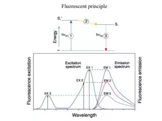
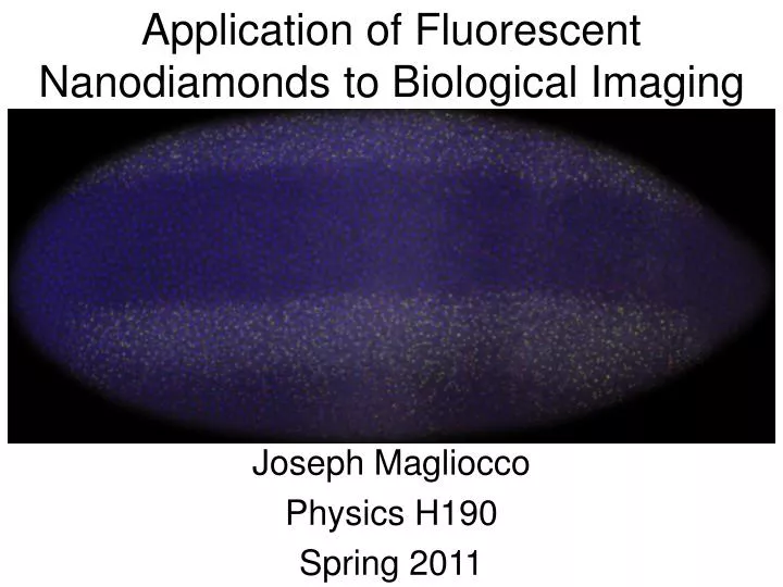
Application of Fluorescent Nanodiamonds to Biological Imaging. Joseph Magliocco Physics H190 Spring 2011. The Basics: Excitation and Emission. Jablonski Diagram. Image from www.icecube.berkeley.edu. The Basics: Excitation and Emission. Spectra. -“Mirror image” rule about ZPL

E N D
Application of Fluorescent Nanodiamonds to Biological Imaging Joseph Magliocco Physics H190 Spring 2011
The Basics: Excitation and Emission Jablonski Diagram Image from www.icecube.berkeley.edu
The Basics: Excitation and Emission Spectra -“Mirror image” rule about ZPL -apply filters for specific excitation and emission wavelengths Image from www.currentprotocols.com
The Basics: Phosphorescence and Photobleaching -Slow emission rates for phosphorescence since the transitions are “not allowed” -Phosphorescence is almost never seen in solution, only in solid state -When a fluorophore is bleached, it is essentially unusable Image from Phys177 Lecture by Carlos Bustamante, Spring 2010
kf Φ = Σki The Basics: Quantum Yield Quantum Yield = number of photons emitted via fluorescence vs. number of photons absorbed ki could be due to fluorescence, internal conversions (phonons), quenching, intersystem crossing (phosphorescence), or any other decay process from the excited state
Two types of applicable FNDs From Nanodiamonds, First Applications in Biology and Nanoscale Medicine by Dean Ho; Chapter 6 – Development and Use of Fluorescent Nanodiamonds as Cellular Markers by Huan-Cheng Chang
Two types of applicable FNDs • Red FNDs (rFNDs) due to NV centers • 140nm rFNDs are mainly NV-, 35nm rFNDs show spectral features of both NV0 and NV-
Two types of applicable FNDs • Green FNDs (gFNDs) due to H3 centers • Both 70nm particles and 140nm particles are predominantly H3, but 140nm shows signs of NV- sideband
Comparison to other fluorophores: Bleaching -gFNDs (red) vs. fluorescent polystyrene nanospheres (blue) -rFNDs (red) vs. fluorescent polystyrene nanospheres (blue) rFNDs exhibited no photobleaching for up to 8 hours while gFNDs exhibited no photobleaching for up to 5 hours.
N-V-N (λem = 531): .95 NV- (λem = 685): .99 Alexa 488 (λem = 519): .92 Alexa 660 (λem = 690): .37 Comparison to Alexa Fluorophores: Quantum Yields FNDs Alexa Fluor Note that Alexa dyes are named after absorption wavelengths Conclusion: FNDs are more efficient and therefore brighter than common Alexa dyes
Other useful properties of FNDs • Cells can take in FNDs (only shown in HeLa cells) • Can conjugate FNDs to proteins, nucleic acids, and carbohydrates • Nontoxic (as opposed to Quantum Dots and other dyes) • Chemically and thermally stable
One potential application: single-particle tracking Motor proteins? DNA translocases?
Future Directions • Cost! • Single vacancy per nanodiamond
References and Acknowledgements • Nanodiamonds, First Applications in Biology and Nanoscale Medicine by Dean Ho; Chapter 6 – Development and Use of Fluorescent Nanodiamonds as Cellular Markers by Huan-Cheng Chang • Physics 177 (Biophysics), Spring 2010, Carlos Bustamante • Alexa Fluorophore website (www.invitrogen.com) • Images cited on slides