Download
1 / 14
140 likes | 197 Views
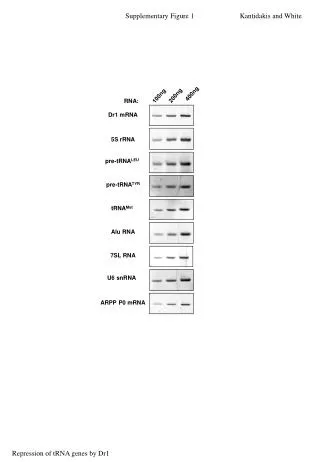
50. mlp1 D -. HXK1. 40. 30. mRNA levels. 20. 10. 0. 1'. 5'. 0'. 1'. 5'. 20'. 40'. 60'. 10'. 20'. 40'. 60'. 10'. 120'. 4.5hr. GLU. GAL induction. GLU repression. mlp1 D -. GAL1::FMP27. 50. 40. mRNA levels. 30. 20. 10. 0. 0'. 1'. 5'. 1'. 5'. 10'. 20'. 40'.

E N D
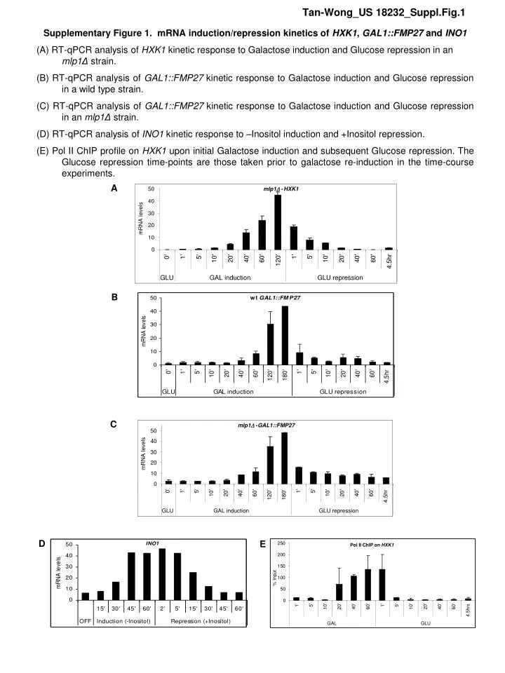
50 mlp1D - HXK1 40 30 mRNA levels 20 10 0 1' 5' 0' 1' 5' 20' 40' 60' 10' 20' 40' 60' 10' 120' 4.5hr GLU GAL induction GLU repression mlp1D - GAL1::FMP27 50 40 mRNA levels 30 20 10 0 0' 1' 5' 1' 5' 10' 20' 40' 60' 10' 20' 40' 60' 120' 180' 4.5hr GLU GAL induction GLU repression 250 Pol II ChIP on HXK1 200 150 % Input 100 50 0 1' 5' 1' 5' 40' 60' 10' 20' 40' 60' 10' 20' 4.5hrs GAL GLU Tan-Wong_US 18232_Suppl.Fig.1 Supplementary Figure 1. mRNA induction/repression kinetics of HXK1, GAL1::FMP27 and INO1 (A) RT-qPCR analysis of HXK1 kinetic response to Galactose induction and Glucose repression in an mlp1Δstrain. (B) RT-qPCR analysis of GAL1::FMP27 kinetic response to Galactose induction and Glucose repression in a wild type strain. (C) RT-qPCR analysis of GAL1::FMP27 kinetic response to Galactose induction and Glucose repression in an mlp1Δstrain. (D) RT-qPCR analysis of INO1 kinetic response to –Inositol induction and +Inositol repression. (E) Pol II ChIP profile on HXK1 upon initial Galactose induction and subsequent Glucose repression. The Glucose repression time-points are those taken prior to galactose re-induction in the time-course experiments. A B C D E
1 2 3 4 (GLU-GAL) (GLU-GAL) (GLU-GAL) (GLU-GAL) (GLU-GAL) (GLU-GAL) (GLU-GAL-GLU 60’-GAL) (GLU-GAL-GLU 60’-GAL) (GLU-GAL-GLU 60’-GAL) (GLU-GAL-GLU 60’-GAL) (GLU-GAL-GLU 60’-GAL) (GLU-GAL-GLU 60’-GAL) (GLU-GAL-GLU 4.5hr-GAL) (GLU-GAL-GLU 4.5hr-GAL) (GLU-GAL-GLU 4.5hr-GAL) (GLU-GAL-GLU 4.5hr-GAL) (GLU-GAL-GLU 4.5hr-GAL) (GLU-GAL-GLU 4.5hr-GAL) A(III) HXK1 primers 1-2 GAL induction 1’ 5’ 10’ 20’ 40’ 60’ 120’ +ve +ve +ve GAL induction GAL induction GAL induction GAL induction GAL induction GAL induction GAL induction GAL induction GAL induction GAL induction GAL induction 1’ 1’ 1’ 1’ 1’ 1’ 1’ 1’ 1’ 1’ 5’ 5’ 5’ 5’ 5’ 5’ 5’ 5’ 5’ 5’ 10’ 10’ 10’ 10’ 10’ 10’ 10’ 10’ 10’ 10’ 20’ 20’ 20’ 20’ 20’ 20’ 20’ 20’ 20’ 20’ 40’ 40’ 40’ 40’ 40’ 40’ 40’ 40’ 40’ 40’ 60’ 60’ 60’ 60’ 60’ 60’ 60’ 60’ 60’ 60’ 120’ 120’ 120’ 120’ 120’ 120’ 120’ 120’ 120’ 120’ 1’ 5’ 10’ 20’ 40’ 60’ 120’ GLU-GAL-GLU 60’-GAL GLU-GAL +ve +ve GLU-GAL-GLU 4.5hr-GAL GLU repression GLU repression GLU repression GLU repression GLU repression GLU repression GLU repression GLU repression GLU repression GLU repression GLU repression GLU repression 1’ 1’ 1’ 1’ 1’ 1’ 1’ 1’ 1’ 1’ 1’ 1’ 5’ 5’ 5’ 5’ 5’ 5’ 5’ 5’ 5’ 5’ 5’ 5’ 10’ 10’ 10’ 10’ 10’ 10’ 10’ 10’ 10’ 10’ 10’ 10’ 20’ 20’ 20’ 20’ 20’ 20’ 20’ 20’ 20’ 20’ 20’ 20’ 40’ 40’ 40’ 40’ 40’ 40’ 40’ 40’ 40’ 40’ 40’ 40’ 60’ 60’ 60’ 60’ 60’ 60’ 60’ 60’ 60’ 60’ 60’ 60’ 4.5hr 4.5hr 4.5hr 4.5hr 4.5hr 4.5hr 4.5hr 4.5hr 4.5hr 4.5hr 4.5hr 4.5hr +ve +ve Tan-Wong_US 18232_Suppl.Fig.2A • Supplementary Figure 2A. HXK1 3C controls for WT and mlp1Δstrains. • The following controls were performed for HXK1 MseI 3C on the WT and mlp1Δ strains – (I) Loading control PCR on HXK1 using the amplicon ‘D’ in Fig1A; (II) Positive control for MseI3C processing on SEN1; (III) 3C negative controls probing promoter-internal fragment interactions (primer combination 1-2) and (IV) promoter-distal fragment interactions (primer combination 1-4) to control for specificity of the gene loop (V) (primers 1-3 as in Fig. 1A). All 3C PCRs were performed alongside a PCR positive control using MseI digested and ligated genomic DNAto control for all possible ligation combinations and primer efficiency. WT mlp1Δ A(I) HXK1 loading control A(II) SEN1 3C control A(IV) HXK1 primers 1-4 A(V) HXK1 primers 1-3
1 3 2 4 (GLU-GAL) (GLU-GAL) (GLU-GAL-GLU 60’-GAL) (GLU-GAL-GLU 60’-GAL) (GLU-GAL-GLU 4.5hr-GAL) (GLU-GAL-GLU 4.5hr-GAL) GAL induction GAL induction GAL induction GAL induction 1’ 1’ 1’ 1’ 5’ 5’ 5’ 5’ 10’ 10’ 10’ 10’ 20’ 20’ 20’ 20’ 40’ 40’ 40’ 40’ 60’ 60’ 60’ 60’ 120’ 120’ 120’ 120’ GLU repression GLU repression GLU repression GLU repression 1’ 1’ 1’ 1’ 5’ 5’ 5’ 5’ 10’ 10’ 10’ 10’ 20’ 20’ 20’ 20’ 40’ 40’ 40’ 40’ 60’ 60’ 60’ 60’ 4.5hr 4.5hr 4.5hr 4.5hr Tan-Wong_US 18232_Suppl.Fig.2B Supplementary Figure 2B. GAL1::FMP27 3C controls. (B) Controls for GAL1::FMP27 EcoRI 3C – (I) Loading control PCR on GAL1::FMP27; (II) Positive control for EcoRI3C processing using FMP27 primers 1-2. Negative controls were performed along FMP27,probing promoter-internal fragment interactions (primer combination 1-3) to control for specificity of the gene loop (primers 1-4 as in Fig. 3A). Data is not shown since, as expected, no 3C signals were obtained and these negative controls have been published previously (1). All 3C PCRs were performed alongside a PCR positive control using EcoRI digested and ligated genomic DNAto control for all possible ligation combinations and primer efficiency. WT mlp1Δ B(I) GAL1::FMP27 loading control B(II) FMP27 primers 1-2
1 2 3 4 WT sua7-1 GLU o/n GAL 60’ GLU 60’ GLU o/n GAL 60’ GLU 60’ + WT sua7-1 GLU o/n GAL 60’ GLU 60’ GLU o/n GAL 60’ GLU 60’ + Tan-Wong_US 18232_Suppl.Fig.2C • Supplementary Figure 2C. HXK1 3C controls for isogenic WT and sua7-1 strains. • The following controls were performed for HXK1 MseI 3C on the isogenic wildtype and sua7-1 strains – (I) Loading control PCR on HXK1 using the amplicon ‘D’ in Fig1A; (II) Positive control for MseI3C processing on SEN1; (III) 3C negative control probing promoter-internal fragment interaction (primer combination 1-2) to control for specificity of the gene loop (primers 1-3 as in Fig. 5A). All 3C PCRs were performed alongside a PCR positive control (‘+’) using MseI digested and ligated genomic DNAto control for all possible ligation combinations and primer efficiency. A(I) A(II) WT sua7-1 GLU o/n GAL 60’ GLU 60’ GLU o/n GAL 60’ GLU 60’ + A(III) HXK1 primers 1-2
I4 I1 (II) I2 – I1 o/n OFF o/n OFF o/n OFF o/n OFF 1hr ON 1hr ON 1hr ON 1hr ON 1hr OFF 1hr OFF 1hr OFF 1hr OFF 1hr ON 1hr ON 1hr ON 1hr ON 12hr OFF 12hr OFF 12hr OFF 12hr OFF 1hr ON 1hr ON 1hr ON 1hr ON + + + + (III) I2 – I4 (IV) I2 – I6 Tan-Wong_US 18232_Suppl.Fig.2D Supplementary Figure 2D. INO1 3C controls. (C) Controls for INO1 HaeIII 3C – (I) Loading control PCR on INO1; (II) Positive control for EcoRI3C processing using INO1 primers I1-I2; (III) 3C negative controlprobing promoter-internal fragment interactions (primer combination I2-I4) to control for specificity of the gene loop (IV; primers I1-I6) as in Fig. 4A). All 3C PCRs were performed alongside a PCR positive control (‘+’) using HaeIII digested and ligated genomic DNAto control for all possible ligation combinations and primer efficiency. (I) Loading control
180 GLU o/n - GAL 1hr 160 GLU-GAL-GLU60'-GAL1hr 140 120 100 mRNA levels 80 60 40 20 0 WT sua7-1 Tan-Wong_US 18232_Suppl.Fig.3 Supplementary Figure 3. sua7-1 re-induction analysis Induction/re-induction time-course data for sua7-1 and its isogenic wildtype, showing a comparison between induction following long-term (overnight) repression (white bars) and re-induction following short-term (60min.) intervening repression. Consistent with the data in Figure 5C, wildtype shows faster re-induction memory, whereas this memory is absent in the sua7-1 mutant.
Tan-Wong_US 18232_Suppl.Fig.4 Supplementary Figure 4. Analysis of GAL:FMP27 long-term induction kinetics GAL1:FMP27 RT-PCR analysis showing long-term mRNA induction profile in a WT strain. The top and bottom graphs are similar, except that the bottom graph contains a wider y-axis scale as it includes the additional 5hr and 8hr time-points. It is observed that stable mRNA levels increase greatly between 3hrs and 5hrs, with the ‘memory’ conditions still providing the highest levels. A drop in mRNA levels is observed between 5hrs and 8hrs, indicating possible transcriptional auto-regulation or mRNA stability regulation at these much later time-points. GLU-GAL GLU-GAL-GLU60’-GAL GLU-GAL-GLU4.5hr-GAL
Galactose re-induction 1hr Galactose re-induction 2hr GAL 1hr 100 GAL 2hr 80 GAL - GLU 1hr - GAL 1hr GAL - GLU 1hr - GAL 2hr mRNA levels 60 40 20 0 WT nup2D Tan-Wong_US 18232_Suppl.Fig.5 Supplementary Figure 5. Nup2 deletionhas no effect on transcriptional memory. GAL1::FMP27RT-qPCR data showing kinetics of Galactose induction and re-induction in wildtype and nup2Δ. Time-points were taken at 1hr (empty box) and 2hrs (grey box) of first-round galactose induction (after overnight growth in glucose) and at 1hr (hashed box) and 2hrs (black box) of second-round galactose re-induction (after an intervening 1hr of glucose repression). The nup2Δ strain shows transcriptional memory effects similar to wildtype, consistent with the 3C data in Fig. 1D showing maintenance of the gene memory-loop in nup2Δ. Galactose 1hr Glucose 1hr O/N Glucose Galactose 2hr
ADH1 control 100 80 ADH1 mRNA levels (normalized to PYK1) 60 40 20 0 WT WT WT WT WT sua7-1 GLU GAL GAL GAL GAL GAL o/n 10' 40' 1hr 2hrs 1hr Tan-Wong_US 18232_Suppl.Fig.6 Supplementary Figure 6. ADH1 transcript levels remain stable during glucose/galactose treatment and between WT and sua7-1 strains Control experiment with ADH1 mRNA normalized to PYK1 transcripts. ADH1 mRNA levels remain largely stable between WT and sua7-1 strains and during glucose/galactose treatments, confirming its reliable use as a normalization control in our experimental conditions for the HXK1 and GAL1:FMP27 RT-qPCR analysis.
Tan-Wong_US 18232_Suppl.Fig.7 Supplementary Figure 7. TBP is maintained at GAL1:FMP27 during 1hr glucose repression in wildtype, but not in mlp1Δ Anti-TBP ChIP performed on wildtype (WT) and mlp1Δstrains at the GAL1::FMP27 promoter at the time-points of galactose growth (GAL) and a subsequent 60min glucose repression (GLU60’). This latter time-point is that at which memory gene loops and transcriptional memory are maintained in wildtype but not in mlp1Δ. As seen here, TBP is still abundantly present during 1hr glucose repression in wildtype, but decreases in an mlp1Δmutant strain in which memory is lost. TBP ChIP WT 2.0 mlp1D 1.5 1.0 0.5 0.0 GAL GLU
Glucose 60’ O/N Glucose Galactose 60’ Glucose 4.5hrs Untagged GAL o/n GAL o/n GLU 60’ GLU 4.5hrs GLU o/n WT mlp1Δ WT mlp1Δ WT mlp1Δ WT mlp1Δ WT mlp1Δ Gal1-GFP (~85kDa) Actin (~42kDa) Tan-Wong_US 18232_Suppl.Fig.8A Supplementary Figure 8A. A difference in Gal1 protein levels is not the cause of the memory loss in mlp1Δmutants. Western blot analysis showing Gal1-GFP and Actin (as loading control). Gal1 protein levels are not detectible in the wildtype and mlp1Δ strains, even during the intervening repression time-point of ‘GLU 60’, during which wildtype cells retain memory, but mlp1Δ cells do not. We presume that the 60’ Galactose induction period prior to short term 60’ Glucose repression or longer term 4.5 hrs repression is insufficient time to accumulate Gal1-GFP protein. These results argue for a Mlp1 (and associated gene-loop) specific memory effect, and against a solely cytoplasmic effect of Gal1 in conferring memory (Zacharioudakis et al, 2007).
Glucose 60’ O/N Glucose Galactose 60’ Glucose 4.5hrs Tan-Wong_US 18232_Suppl.Fig.8B Supplementary Figure 8B. FACS analysis at representative time points showing single cell analysis of Gal1-GFP fluorescence measurements during repression time points. Gal1-GFP-tagged yeast strains expressing GFP fluorescence byFACs analysis shows similar fluorescence levels between wild type and mlp1Δ strains. i) Diagram of fluorescence measurement (Y-axis: FL1-H) against 30000 counts of yeast cells (X-axis: SSC-H). Left panel is the untagged strain (untag) while right panel shows the fluorescence of Gal1-GFP-tagged yeast grown in galactose overnight (Gal o/n). ii) Histogram of FACS analysis showing mean Gal1-GFP fluorescence values, over 3 biological repeats, at representative time points taken from wild type and mlp1∆ grown continuously in glucose o/n (Glu o/n), washed in water before activating in galactose media for 60 minutes. The strains were then subjected to repression in glucose media for 60 minutes (60’ Glu) and and 4.5 hours (4.5hr Glu). Note that as for the western blot analysis (Supplemenary Figure 8A) no fluorescent cells were detected during the short Glucose repression period (memory) arguing against Gal1-GFP effects in this process. i) Gal o/n untag ii) wild type 100 mlp1 ∆ 50 % population of flourescence -1 untag Glu o/n 60’ Glu 4.5 hr Glu Gal o/n
Tan-Wong_US 18232_Suppl.Fig.9 Supplementary Figure 9. Western blot control of Mlp1-GFP tagged WT and sua7-1 strains Western blot control of isogenic wildtype and sua7-1 strains (3 clones each shown here) that were Mlp1-GFP tagged using homologous recombination. The western blot has been probed with α-GFP and α –Actin (loading control). A Gal1-GFP tagged strain was used as a control for the GFP antibody; a Mlp1-GFP strain from Feuerbach et al (2003) was used as a positive control; and an untagged strain was used as a negative control.
Tan-Wong_US 18232_Suppl.Table.1 Supplementary Table 1. Primers used in ChIP, 3C and RT-qPCR analysis