Download
1 / 29
290 likes | 452 Views
Chapter 42: Circulation and Gas Exchange. Ms. Klinkhachorn April 29, 2011 AP Biology. The need for a circulatory system. Need to exchange gases, nutrients, and wastes BUT Diffusion is slow and only occurs over small distances How do we get around this?. Two Solutions in Nature.

E N D
Chapter 42: Circulation and Gas Exchange Ms. Klinkhachorn April 29, 2011 AP Biology
The need for a circulatory system • Need to exchange gases, nutrients, and wastes • BUT Diffusion is slow and only occurs over small distances • How do we get around this?
Two Solutions in Nature • Organisms with body shapes and sizes that keep almost all of their cells in contact with the environment • Can have a gastrovascular cavity that help a dual-function (digestion and distribution)
Two Solutions in Nature • Organism has a circulatory system • More complex species • Circulatory systems have 3 parts: • Blood (circulatory fluid) • Vessels (tubes that move the fluid) • Heart (structure that pumps the fluid)
Main Blood Vessel Types • Arteries • Carry blood away from the heart • Capillaries • Microscopic vessels (one cell layer thick) • Sites of diffusion • Veins • Carry blood back to the heart
Parts of the Heart • Atria (atrium) are heart chambers that receive blood from veins • Ventricles take blood from the atria and then pump the blood back out
Circulatory Systems Can Vary • Vary based on the organism • Mammals and birds have hearts with 4 chambers • Reptiles and amphibians have 3 • Fish have 2 • In mammals, left side of the heart deals with oxygen-rich blood while right side deals with oxygen-poor blood
Amphibians Reptiles (Except Birds) Mammals and Birds Lung and skin capillaries Lung capillaries Lung capillaries Fig. 42-5 Right systemic aorta Pulmocutaneous circuit Pulmonary circuit Pulmonary circuit Atrium (A) Atrium (A) A A A A V V Ventricle (V) V V Left systemic aorta Left Right Left Right Right Left Systemic circuit Systemic circuit Systemic capillaries Systemic capillaries Systemic capillaries
Capillaries of head and forelimbs Superior vena cava 7 Pulmonary artery Pulmonary artery Capillaries of right lung Aorta 9 Capillaries of left lung Fig. 42-6 3 3 2 4 11 Pulmonary vein Pulmonary vein 5 1 Right atrium Left atrium 10 Right ventricle Left ventricle Inferior vena cava Aorta Capillaries of abdominal organs and hind limbs 8
Steps of Circulation in Mammals • Blood is pumped from right ventricle to the lungs via the pulmonary artery • Blood picks up oxygen in capillary beds of lungs and releases carbon dioxide • Blood returns to the left atrium via the pulmonary vein and move into the left ventricle • Blood moves through the aorta and ultimately to other places in the body
Steps of Circulation in Mammals • Blood travels through body and, in capillary beds, releases oxygen and picks up carbon dioxide • Blood returns back to the right atrium of the heart via the vena cava • Blood flows into the right ventricle and the cycle restarts
Fig. 42-7 Pulmonary artery Aorta Pulmonary artery Right atrium Left atrium Semilunar valve Semilunar valve Atrioventricular valve Atrioventricular valve Right ventricle Left ventricle
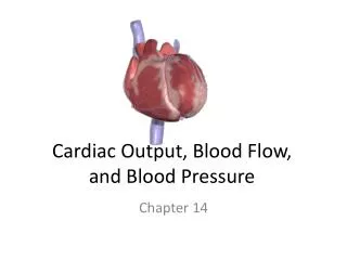
Cardiac Cycle • Cycle of contraction and relaxation = cardiac cycle (time from one beat to the next) • Systole: contraction (pumping) phase • Diastole: relaxation (filling) phase • Heart rate = beats per minute • SA node is the pacemaker of the heart • This controls the heart contractions
Blood Pressure • Typical blood pressure for a 20 year old at rest is 120/70 • First number is the systolic pressure • Second number is the diastolic pressure
Blood Components • Suspended in blood plasma are two types of cells: • Red blood cells (erythrocytes) transport oxygen via hemoglobin • White blood cells (leukocytes) function as defense in the immune system • Platelets are fragments of cells that are involved in clotting
Fig. 42-17 Plasma 55% Constituent Major functions Cellular elements 45% Cell type Number per µL (mm3) of blood Functions Solvent for carrying other substances Water Erythrocytes (red blood cells) Transport oxygen and help transport carbon dioxide 5–6 million Ions (blood electrolytes) Sodium Potassium Calcium Magnesium Chloride Bicarbonate Osmotic balance, pH buffering, and regulation of membrane permeability Separated blood elements Leukocytes (white blood cells) Defense and immunity 5,000–10,000 Plasma proteins Albumin Osmotic balance pH buffering Lymphocyte Basophil Fibrinogen Clotting Eosinophil Immunoglobulins (antibodies) Defense Neutrophil Monocyte Substances transported by blood Nutrients (such as glucose, fatty acids, vitamins) Waste products of metabolism Respiratory gases (O2 and CO2) Hormones 250,000– 400,000 Platelets Blood clotting
RBC Structure and Function • Disc shaped • Maximize surface area for oxygen to bind to hemoglobin • Lack nuclei • Gives more space • Lack mitochondria • Oxygen held isn’t used
Fig. 42-18-4 Red blood cell Collagen fibers Platelet plug Fibrin clot Platelet releases chemicals that make nearby platelets sticky Clotting factors from: Platelets Damaged cells Plasma (factors include calcium, vitamin K) Prothrombin Thrombin Fibrinogen Fibrin 5 µm
Fig. 42-20 Smooth muscle Connective tissue Plaque Endothelium (a) Normal artery (b) Partly clogged artery 50 µm 250 µm
Fig. 42-20a Smooth muscle Connective tissue Endothelium 50 µm (a) Normal artery
Fig. 42-20b Plaque (b) Partly clogged artery 250 µm
Countercurrent Exchange • Gills in fish • Water flow through, opposite the direction of blood flow • Picks up oxygen from the water
Fig. 42-21a Parapodium (functions as gill) (a) Marine worm
Fig. 42-21b Gills (b) Crayfish
Fig. 42-21c Coelom Gills Tube foot (c) Sea star
Fig. 42-22 Fluid flow through gill filament Oxygen-poor blood Anatomy of gills Oxygen-rich blood Gill arch Lamella Gill arch Gill filament organization Blood vessels Water flow Operculum Water flow between lamellae Blood flow through capillaries in lamella Countercurrent exchange PO2 (mm Hg) in water 150 120 90 60 30 Gill filaments Net diffu- sion of O2 from water to blood 110 80 50 20 140 PO2 (mm Hg) in blood
Flow of Oxygen • Into your mouth or nose • Past the voice box (larynx) • Into the trachea (windpipe) • Into one of the two bronchi • Into a branch called a bronchiole • Into an alveoli
Fig. 42-24 Branch of pulmonary vein (oxygen-rich blood) Branch of pulmonary artery (oxygen-poor blood) Terminal bronchiole Nasal cavity Pharynx Larynx Alveoli (Esophagus) Left lung Trachea Right lung Bronchus Bronchiole Diaphragm Heart SEM Colorized SEM 50 µm 50 µm