Download
1 / 29
340 likes | 694 Views
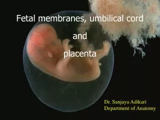
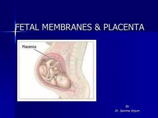
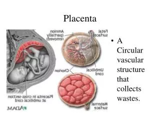
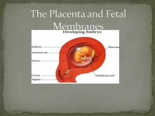
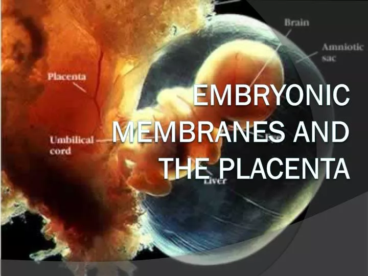
Embryonic Membranes and the Placenta. Placenta and Fetal Membranes. Amnion - Epiblast / Extraembryonic Mesoderm Yolk Sac - Hypoblast / Extraembryonic Mesoderm Allantois - Embryonic Hindgut Chorion - Trophoblasts / Extraembryonic Mesoderm Placenta - Chorion / Maternal Decidua.

E N D
Placenta and Fetal Membranes • Amnion - Epiblast / Extraembryonic Mesoderm • Yolk Sac - Hypoblast / Extraembryonic Mesoderm • Allantois - Embryonic Hindgut • Chorion - Trophoblasts / Extraembryonic Mesoderm • Placenta - Chorion / Maternal Decidua
Amnion • Amnionicmembrane is two cell layers • As the amnion enlarges it encompasses the embryo • Amnion forms the epithelial layer of the umbilical cord • With embryo growth the amnion obliterates the chorionic cavity • Amnionic sac is fluid filled called amnionic fluid: the embryo is bathed in the fluid
Extraembryonic Tissues • 8 days • 9 days 9 days • 14days
Amniotic Fluid • Up to week 20 - fluid is similar to fetal serum (keratinization) • After 20 weeks – Contribution from urine, maternal serum filtered • thru endothelium of nearby vessels, filtration from fetal • vessels in cord • Near birth - can contain fetal feces called meconium • Near birth – amnionic fluid (500-1000 ml) exchanges every 3 hrs • 1) across the amnion – exchange with maternal fluids. • 2) fetal swallowing (20 ml/hour) – to gut – adsorption by • fetus – out the umbilical cord to placenta. • Hydraminos – Excess fluid (>2000 ml), esophageal atresia • Oligohydramnios – Insufficient fluid (<500 ml), renal agenesis
Amnion Function • Mechanical protection: hydrostatic pressure • Allows free movement - which aids in neuromuscular • development • Antibacterial • Allow for fetal growth • Protection from adhesions
Yolk Sac • Hypoblast - the primary yolk sac or Heuser's membrane. • Day 12 - Second wave of cell migration - forms definitive yolk sac • Early nutrition (2-3 weeks) for the embryo - later shrinking • Derivatives: • Primordial germ cells • The early gut, epithelium of the respiratory and digestive tracts
Allantois • Endodermalorigin – caudal outpocketing of the yolk sac • Involved in early hematopoiesis • The allantois blood vessels - artery and vein – becomes the umbilical vessels • Remnants of Allantois becomes the urachus ligament that connects the belly button to the bladder
Chorion • Chorionic cavity (extraembryoniccoelom)- lined with extraembryonicmesoderm • The Chorion / maternal endometrium forms the placenta • Chorion forms stem villi
Stem Villi • Chorionic Plate – Stem villi extends from this tissue • Primary stem villi (day 11-13) - finger-like protrusions • into endometrium– • Secondary stem villi (day 16) - extraembryonic mesoderm invasion into villi core. • Tertiary stem villus (21 day) - extraembryonic vessels • chorionic arteries and veins derived from extraembryonic mesoderm.
Stem Villi • Cytotrophoblasticshell – surrounds embryo; direct contact with maternal decidual cells • Anchoring Villi– give off cytotrophoblastic extensions - • anchoring because they represent the real maternal embryo link • Floating Villi– branches off anchoring villi – dangles freely in maternal blood
Making the Placenta • By 8 weeks - chorionic stem villi over the entire surface of the chorionic sac • Those villiincrease in size and more villi form. • The villi continue to enlarge during most of gestation. • Endometrial vessels - spiral arteries and endometrial veins form
Umbilical Cord • One umbilical vein, • Two umbilical arteries • Wharton’s jelly – • connective tissue • surrounding vessels • Allantois • Yolk Stalk (vitelline duct)
Placenta as an Endocrine Organ • Human Chorionic Gonadotropin– Corpus Luteum (declines after 8 weeks) • Progesterone – High levels by the end of first trimester • Estrogen – Synthesis involves enzymatic activity of fetal adrenal gland and liver • Chorionic Somatomammotropin– Human Placental Lactogen– similar to GH (growth, lactation, lipid and carbohydrate metabolism) • Placental Growth Hormone – similar to GH – Replaces maternal GH by 15 wks – enhances blood glucose levels