1 / 41

Brain Imaging

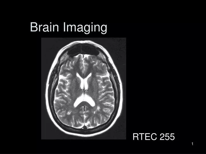
Brain Imaging. RTEC 255. Brain Stem. Skull fractures with rupture of the meningeal arteries can cause epidural hematoma. Brain trauma with rupture of the meningeal veins (subdural space) can cause subdural hematoma. Falx Cerebelli separates the two cerebellar hemispheres.

tuan
Download Presentation

Brain Imaging

An Image/Link below is provided (as is) to download presentation Download Policy: Content on the Website is provided to you AS IS for your information and personal use and may not be sold / licensed / shared on other websites without getting consent from its author. Content is provided to you AS IS for your information and personal use only. Download presentation by click this link. While downloading, if for some reason you are not able to download a presentation, the publisher may have deleted the file from their server. During download, if you can't get a presentation, the file might be deleted by the publisher.

E N D

Presentation Transcript


  1. Brain Imaging RTEC 255

  2. Brain Stem

  3. Skull fractures with rupture of the meningeal arteries can cause epidural hematoma

  4. Brain trauma with rupture of the meningeal veins (subdural space) can cause subdural hematoma

  5. Falx Cerebelli separates the two cerebellar hemispheres

  6. Tentorium Cerebelli which spreads out like a tent, forms a partition between the cerebrum & cerebellum

  7. Ventricle Anatomy

  8. Choroid Plexus

  9. Cisterns – widended subarachnoid space

  10. Basal Nuclei or Basal Ganglia • Collection of subcortical gray matter. • Collectively, contribute to the planning and programming of muscle action and movement. Serve as a relay station between the thalamus and the cerebral cortex.

  11. Brain Vascular Anatomy

  12. Circle of Willis

  13. Circle of Willis

  14. Cerebral Angiogram

  15. MRI - MRA

  16. Questions ?

More Related