Download
1 / 32
360 likes | 698 Views
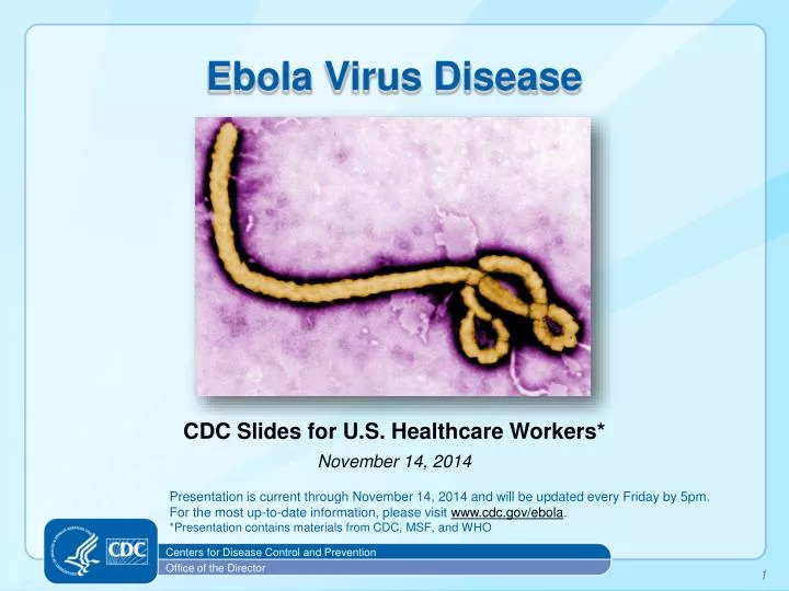
Ebola Virus Disease. CDC Slides for U.S. Healthcare Workers* November 14, 2014. P resentation is current through November 14, 2014 and will be updated every Friday by 5pm. For the most up-to-date information, please visit www.cdc.gov/ebola .

E N D
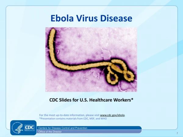
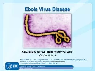
Ebola Virus Disease CDC Slides for U.S. Healthcare Workers* November 14, 2014 Presentation is current through November 14, 2014 and will be updated every Friday by 5pm. For the most up-to-date information, please visit www.cdc.gov/ebola. *Presentation contains materials from CDC, MSF, and WHO Centers for Disease Control and Prevention Office of the Director 1
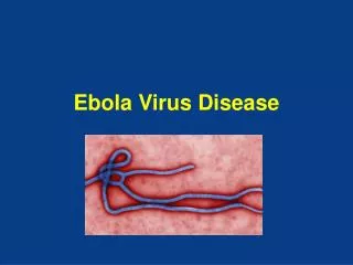
Ebola Virus • Prototype Viral Hemorrhagic Fever Pathogen • Filovirus: enveloped, non-segmented, negative-stranded RNA virus • Severe disease with high case fatality • Absence of specific treatment or vaccine • >20 previous Ebola and Marburg virus outbreaks • 2014 West Africa Ebola outbreak caused by Zaire ebolavirusspecies (five known Ebola virus species) 2
Ebola Virus • Zoonotic virus – bats the most likely reservoir, although species unknown • Spillover event from infected wild animals (e.g., fruit bats, monkey, duiker) to humans, followed by human-human transmission 3
Figure. Ebola virus disease (EVD) cumulative incidence* — West Africa, October 18, 2014 * Cumulative number of reported EVD cases per 100,000 persons since December 22, 2013. MMWR 2014;63(43):978-981 4
2014 Ebola Outbreak, West Africa WHO Ebola Response Team. N Engl J Med 2014. DOI: 10.1056/NEJMoa1411100http://www.nejm.org/doi/full/10.1056/NEJMoa1411100?query=featured_ebola#t=articleResults 5
EVD Cases and Deaths* Updated case counts available at http://www.cdc.gov/vhf/ebola/outbreaks/2014-west-africa/case-counts.html. *Reported by WHO using data from Ministries of Health **The outbreaks of EVD in Senegal and Nigeria were declared over on October 17 and 19, respectively. 6
EVD Cases (United States) • EVD has been diagnosed in the United States in four people, one (the index patient) who traveled to Dallas, Texas from Liberia, two healthcare workers who cared for the index patient, and one medical aid worker who traveled to New York City from Guinea • Index patient – Symptoms developed on September 24, 2014 approximately four days after arrival, sought medical care at Texas Health Presbyterian Hospital of Dallas on September 26, was admitted to hospital on September 28, testing confirmed EVD on September 30, patient died October 8. • TX Healthcare Worker, Case 2 – Cared for index patient, was self-monitoring and presented to hospital reporting low-grade fever, diagnosed with EVD on October 10, recovered and released from NIH Clinical Center October 24. • TX Healthcare Worker, Case 3 – Cared for index patient, was self-monitoring and reported low-grade fever, diagnosed with EVD on October 15, recovered and released from Emory University Hospital in Atlanta October 28. • NY Medical Aid Worker, Case 4 – Worked with Ebola patients in Guinea, was self-monitoring and reported fever, diagnosed with EVD on October 24, recovered and released from Bellevue Hospital in New York City November 11. Information on U.S. EVD cases available at http://www.cdc.gov/vhf/ebola/outbreaks/2014-west-africa/united-states-imported-case.html. 7
EVD Cases (United States) • As of October 31, 2014, four U.S. health workers and one journalist who were infected with Ebola virus in West Africa were transported to hospitals in the United States for care • All the patients have recovered and have been released from the hospital after laboratory testing confirmed that they no longer have Ebola virus in their blood 8
Ebola Virus Transmission • Virus present in high quantity in blood, body fluids, and excreta of symptomatic EVD-infected patients • Opportunities for human-to-human transmission • Direct contact (through broken skin or unprotected mucous membranes) with an EVD-infected patient’s blood or body fluids • Sharps injury (with EVD-contaminated needle or other sharp) • Direct contact with the corpse of a person who died of EVD • Indirect contact with an EVD-infected patient’s blood or body fluids via a contaminated object (soiled linens or used utensils) • Ebola can also be transmitted via contact with blood, fluids, or meat of an infected animal • Limited evidence that dogs become infected with Ebola virus • No reports of dogs or cats becoming sick with or transmitting Ebola 9
Detection of Ebola Virus in Different Human Body Fluids over Time 10
Human-to-Human Transmission • Infected persons are not contagious until onset of symptoms • Infectiousness of body fluids (e.g., viral load) increases as patient becomes more ill • Remains from deceased infected persons are highly infectious • Human-to-human transmission of Ebola virus via inhalation (aerosols) has not been demonstrated 11
EVD Risk Assessment **CDC Website to check current affected areas: www.cdc.gov/vhf/ebola
Ebola Virus Pathogenesis • Direct infection of tissues • Immune dysregulation • Hypovolemia and vascular collapse • Electrolyte abnormalities • Multi-organ failure, septic shock • Disseminated intravascular coagulation (DIC) and coagulopathy Lancet. Mar 5, 2011; 377(9768): 849–862. 13
Early Clinical Presentation • Acute onset; typically 8–10 days after exposure (range 2–21 days) • Signs and symptoms • Initial: Fever, chills, myalgias, malaise, anorexia • After 5 days: GI symptoms, such as nausea, vomiting, watery diarrhea, abdominal pain • Other: Headache, conjunctivitis, hiccups, rash, chest pain, shortness of breath, confusion, seizures • Hemorrhagic symptoms in 18% of cases • Other possible infectious causes of symptoms • Malaria, typhoid fever, meningococcemia, Lassa fever and other bacterial infections (e.g., pneumonia) – all very common in Africa 14
Clinical Features • Nonspecific early symptoms progress to: • Hypovolemic shock and multi-organ failure • Hemorrhagic disease • Death • Non-fatal cases typically improve 6–11 days after symptoms onset • Fatal disease associated with more severe early symptoms • Fatality rates of 70% have been reported in rural Africa • Intensive care, especially early intravenous and electrolyte management, may increase the survival rate 15
Clinical Manifestations by Organ Systemin West African Ebola Outbreak WHO Ebola Response team. NEJM. 2014 16
Examples of Hemorrhagic Signs Hematemesis Gingival bleeding Bleeding at IV Site 17
Laboratory Findings • Thrombocytopenia (50,000–100,000/mL range) • Leukopenia followed by neutrophilia • Transaminase elevation: elevation serum aspartate amino-transferase (AST) > alanine transferase (ALT) • Electrolyte abnormalities from fluid shifts • Coagulation: PT and PTT prolonged • Renal: proteinuria, increased creatinine 18
EVD: Expected diagnostic test results over time Critical information: Date of onset of fever/symptoms IgM IgG viremia RT-PCR 0 3 10 days post onset of symptoms Fever ELISA IgM ELISA IgG IgM: up to 3 – 6 months IgG: 3 – 5 years or more (life-long persistance?) 19
Ebola Virus Diagnosis • Real Time PCR (RT-PCR) • Used to diagnose acute infection • More sensitive than antigen detection ELISA • Identification of specific viral genetic fragments • Performed in select CLIA-certified laboratories • RT-PCR sample collection • Volume: minimum volume of 4mL whole blood • Plastic collection tubes (not glass or heparinized tubes) • Whole blood preserved with EDTA is preferred • Whole blood preserved with sodium polyanetholsulfonate (SPS), citrate, or with clot activator is acceptable 20
Other Ebola Virus Diagnostics • Virus isolation • Requires Biosafety Level 4 laboratory; • Can take several days • Immunohistochemical staining and histopathology • On collected tissue or dead wild animals; localizes viral antigen • Serologic testing for IgM and IgG antibodies (ELISA) • Detection of viral antibodies in specimens, such as blood, serum, or tissue suspensions • Monitor the immune response in confirmed EVD patients 21
Laboratories • CDC has developed interim guidance for U.S. laboratory workers and other healthcare personnel who collect or handle specimens • This guidance includes information about the appropriate steps for collecting, transporting, and testing specimens from patients who are suspected to be infected with Ebola • Specimens should NOT be shipped to CDC without consultation with CDC and local/state health departments Information available at: http://www.cdc.gov/vhf/ebola/hcp/interim-guidance-specimen-collection-submission-patients-suspected-infection-ebola.html 22
Packaging & Shipping Clinical Specimens to CDC for Ebola Testing http://www.cdc.gov/vhf/ebola/hcp/packaging-diagram.html 23
Interpreting Negative Ebola RT-PCR Result • If symptoms started ≥3 days before the negative result • EVD is unlikely consider other diagnoses • Infection control precautions for EVD can be discontinued unless clinical suspicion for EVD persists • If symptoms started <3 days before the negative RT-PCR result • Interpret result with caution • Repeat the test at ≥72 hours after onset of symptoms • Keep in isolation as a suspected case until a repeat RT-PCR ≥72 hours after onset of symptoms is negative 24
Reference: Fowler RA et al. Am J RespirCrit Care Med. 2014 Clinical Management of EVD: Supportive, but Aggressive • Hypovolemia and sepsis physiology • Aggressive intravenous fluid resuscitation • Hemodynamic support and critical care management if necessary • Electrolyte and acid-base abnormalities • Aggressive electrolyte repletion • Correction of acid-base derangements • Symptomatic management of fever and gastrointestinal symptoms • Avoid NSAIDS • Multisystem organ failure can develop and may require • Oxygenation and mechanical ventilation • Correction of severe coagulopathy • Renal replacement therapy 25
Investigational Therapies for EVD Patients • No approved Ebola-specific prophylaxis or treatment • Ribavirin has no in-vitro or in-vivo effect on Ebola virus • Therapeutics in development with limited human clinical trial data • Convalescent serum • Therapeutic medications • Zmapp– chimeric human-mouse monoclonal antibodies • Tekmira– lipid nanoparticle small interfering RNA • Brincidofovir– oral nucleotide analogue with antiviral activity • Vaccines – in clinical trials • Chimpanzee-derived adenovirus with an Ebola virus gene inserted • Attenuated vesicular stomatitis virus with an Ebola virus gene inserted References: 1Huggins, JW et al. Rev Infect Dis 1989; 2Ignatyev, G et al. J Biotechnol 2000; 3Jarhling, P et al. JID 2007 S400; 4Mupapa, K et al. JID 1999 S18; 5Olinger, GG et al. PNAS 2012; 6Dye, JM et al. PNAS 2012; 7Qiu, X et al. Sci Transl Med 2013; 8Qiu, X et al. Nature 2014; 9Geisbert, TW et al. JID 2007; 10Geisbert, TW et al. Lancet 2010; 11Kobinger, GP et al. Virology 2006; 12Wang, D JV 2006; 13Geisbert, TW et al. JID 2011; and 14Gunther et al. JID 2011. 26
Patient Recovery • Case-fatality rate 71% in the 2014 Ebola outbreak • Case-fatality rate is likely much lower with access to intensive care • Patients who survive often have signs of clinical improvement by the second week of illness • Associated with the development of virus-specific antibodies • Antibody with neutralizing activity against Ebola persists greater than 12 years after infection • Prolonged convalescence • Includes arthralgia, myalgia, abdominal pain, extreme fatigue, and anorexia; many symptoms resolve by 21 months • Significant arthralgia and myalgia may persist for >21 months • Skin sloughing and hair loss has also been reported References: 1WHO Ebola Response Team. NEJM 2014; 2Feldman H & Geisbert TW. Lancet 2011; 3Ksiazek TG et al. JID 1999; 4Sanchez A et al. J Virol 2004; 5Sobarzo A et al. NEJM 2013; and 6Rowe AK et al. JID 1999. 27
Practical Considerations for Evaluating Patients for EVD in the United States • CDC encourages all U.S. healthcare providers to • Ask patients with symptoms about a history of travel to West Africa in the 21 days before illness onset • Know the signs and symptoms of EVD • Know the initial steps to take if a diagnosis of EVD is suspected • CDC has developed documents to facilitate these evaluations • The EVD algorithm for the evaluation of a returned traveler • Available at http://www.cdc.gov/vhf/ebola/pdf/ebola-algorithm.pdf • The checklist for evaluation of a patient being evaluated for EVD • Available at http://www.cdc.gov/vhf/ebola/pdf/checklist-patients-evaluated-us-evd.pdf 28
EVD Algorithm for Evaluation of theReturned Traveler **CDC Website to check current affected areas: www.cdc.gov/vhf/ebola Algorithm available at http://www.cdc.gov/vhf/ebola/pdf/ebola-algorithm.pdf Checklist available at http://www.cdc.gov/vhf/ebola/pdf/checklist-patients-evaluated-us-evd.pdf 29
Interim Guidance for Monitoring and Movement of Persons with EVD Exposure • CDC has created guidance for monitoring people exposed to Ebola virus but without symptoms • www.cdc.gov/vhf/ebola/hcp/monitoring-and-movement-of-persons-with-exposure.html
EVD Summary • The 2014 Ebola outbreak in West Africa is the largest in history and has affected multiple countries • Think Ebola: U.S. healthcare providers should be aware of clinical presentation and risk factors for EVD • Human-to-human transmission by direct contact • No human-to-human transmission via inhalation (aerosols) • No transmission before symptom onset • Early case identification, isolation, treatment and effective infection control are essential to prevent Ebola transmission 31
Centers for Disease Control and Prevention Office of the Director 32