Download
1 / 24
240 likes | 339 Views
What do you know & want to know about the Nervous System?. Collins type 1, ~10 lines, sentences related to the question please. Nervous System. Pages 228-237. Functions of the nervous system. 1. Sensory input—gathering info Monitor changes occurring inside & outside the body 2. Integration

E N D
What do you know & want to know about the Nervous System? Collins type 1, ~10 lines, sentences related to the question please
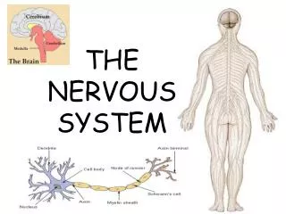
Nervous System Pages 228-237
Functions of the nervous system 1. Sensory input—gathering info • Monitor changes occurring inside & outside the body 2. Integration • Make sense of information &decide if/what action is needed 3. Motor output • The “response” • activates muscles or glands in response to the stimulus
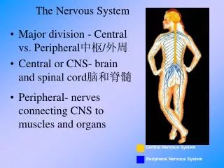
Structural Classification of the NS • Central nervous system (CNS) • Brain • Spinal cord • Peripheral nervous system (PNS) • Nerves outside the brain and spinal cord • Spinal nerves • Cranial nerves
Functional Classification of the Peripheral NS • Sensory (afferent) division • Nerve fibers that carry information to the CNS • “On ramp” • Motor (efferent) division • Nerve fibers that carry impulses away from the CNS • Two subdivisions • Somatic nervous system = voluntary • Autonomic nervous system = involuntary
Practice… Do you have the gist? • In the scenario identify the sensory input, motor effect, and integration (not specifically stated). Also identify the role of the efferent and afferent divisions of the nervous system and explain which specific efferent/motor division is being used. • Fritz was putting a tray of cookies into the oven. The top of his hand accidently grazed the side of the oven and he quickly retracted his hand out of the oven.
Supporting cells of the CNS • Called neuroglia, glia, or glial cells • Literally mean “nerve glue” • supporting cells of the CNS that help protect, support, and insulate • 4 types to know • Astrocytes • Star-shaped • Make up ~½ of the neural tissue in body • Anchor neurons to blood supply • Form a living, protective barrier barrier between capillaries and neurons
Neuroglia AKA Glial Cells 2. Microglia • Spider-like macrophages that destroy debris bacteria and dead brain cells 3. Ependymal Cells • Line the cavities of the brain & spinal cord • Have cilia that move and help circulate cerebrospinal fluid
Neuroglia AKA Glial Cells 4. Oligodendrocytes • Have flat extensions that wrap around nerve fibers • Make myelin sheaths fatty cover that insulates nerve fibers • Glial cells VS. Neurons • Look similar BUT glial cells can’t transmit or conduct nerve impulsesk • Glial cells can divide, neurons can’t (most brain tunmors are gliomas formed by glial cells)
Supporting Cells of the PNS • Schwann Cells • Form myelin sheaths around nerve fibers • MS attacks myelin sheaths, converts them to hard covers that can’t conduct electrical impulses slurred speech, loss of balance, impaired vision, etc • Sattellite Cells • Serve as protective cushioning cells
Something to think about… • What is a Schwann cell? • What part of the NS does it support? • What structural part of the neuron can it form?
Neuron Anatomy • Cell body/soma • Metabolic center • Dendrites • Can have a few or 60 • Receive messages from other neurons • Axon • Can only have ONE • Sends the message from the body
Neuron Anatomy • Axon hillock (where AP is generated) and axon terminal (branches at the end of an axon) • Myelin sheath • Not present on every axon • Pain receptors don’t have myelin on their axons • Help message travel faster • By schwann or oligodendrocytes
Neuron Physiology • Neurons are selectively permeable • Contain ion channels • Voltage gated ion channels only open when there is a change in electrical charge necessary for transmission of an action potential! • At resting state the neurons is polarized (has a slightly negative charge compared to the outside of the neuron) • b/c inside of cell has proteins that have – charges • Has K+ but not enough to balance out the – • At resting state the area around a neuron is more positive
Making an Action Potential • A stimulus causes Na+ to flow into the neuron • If enough Na+ flows in, the neuron’s charge becomes more positive and becomes depolarized (not as negative) causing the nerve to send an electrical signal action potential AKA nerve impulse • Sodium voltage-gated ion channels open and Na+ rushes in make the inside very +
Making an Action Potential • Once one area is positive, the positive charge moves down the axon causing more Na+ channels to open • After Na+ enters, the ions channels for Na+ close but the inside is still very + • Sodium-potassium pumps then use ATP to move Na+ out of the neuron and K+ in to return the charges inside and outside of the neuron to “normal” repolarization • All or Nothing Response • 1 action potential at a time while neuron is recovering it is in a refractory period no nerve impulses an be generated
Action Potentials in Myelinated vs. Not fibers • Myelinated fibers carry messages faster • Use saltatory conduction (charge jumps from node of Ranvier to node of Ranvier) • Non-myelinated fibers transmit messages slower b/c all of the ion channels must open and close to propagate the electrical signal http://184.171.162.94/images/prevjhy.php?u=Oi8vd3d3LnlvdXR1YmUuY29tL3dhdGNoP3Y9REplM18zWHNCT2c%3D&b=5
Animations of AP Generation • http://184.171.162.94/images/prevjhy.php?u=Oi8vd3d3LnlvdXR1YmUuY29tL3dhdGNoP3Y9aWZEMVlHMDdmQjg%3D&b=5 • http://highered.mcgraw-hill.com/sites/0072495855/student_view0/chapter14/animation__the_nerve_impulse.html
Practice • In groups of 2-4 create an analogy that parallels the event that take place to create an action potential/nerve impulse • Consider • the initial locations of Na+ and K+ • the movement of Na+ while the action potential is being created • The movement of K+ immediately after the AP has been generated • How the levels of Na+ and K+ inside and outside of the cell is corrected after the AP has been transmitted
Step By Step… 1. A neuron is more negative inside the cell relative to the outside (polarized) • 2. A stimulus makes causes the cell’s charge to reach the threshold • 3. Na+ channels open and sodium floods into the cell in one section of the axon • 4. The Na+ channels in that area close but the region down the axon gets positive enough to reach threshold Na+ channels open and sodium rushes in… this continues down the axon • 5. The K+ channels open and potassium diffuses out • 6.The cell becomes repolarized BUT K+ is concentrated outside and Na+ is concentrated inside… must swap! • 7. The sodium-potassium pumps move Na+ our of the neuron and K+ into the neuron