Download
1 / 1
10 likes | 143 Views
Trunk Muscles Respond To Task Specific Fatigue In An Opposite Manner As Appendicular Muscles James S. Thomas 1,2,3 , Andrew J. Ross 1 , Brian C. Clark 2,3 , Jeffery Cowen 1 , Richard Pickett 1 , Mathew Linsenmayer 1

E N D
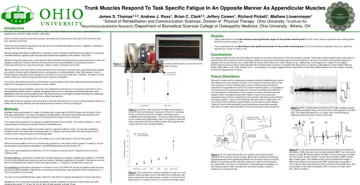
Trunk Muscles Respond To Task Specific Fatigue In An Opposite Manner As Appendicular Muscles James S. Thomas1,2,3, Andrew J. Ross1, Brian C. Clark2,3, Jeffery Cowen1, Richard Pickett1, Mathew Linsenmayer1 1School of Rehabilitation and Communication Sciences, Division of Physical Therapy, Ohio University, 2Institute for Neuromusculoskeletal Research,3Department of Biomedical Sciences College of Osteopathic Medicine, Ohio University, Athens, OH • Introduction • The most common type of pain reported by adults in the United States is low back pain, causing dysfunction, disability and a decline in daily activities. (Day, Nitz) • After the initial incident of acute low back pain, the likelihood of recurrent low back pain is 25% within the first year. (Stanton, Henschke) • There are many treatment approaches for low back pain including therapeutic exercise, modalities, mobilization, manipulation and muscle energy. • Muscle energy techniques are defined as a voluntary muscle contraction performed by the patient “in a precisely controlled direction, against a distinctly executed counter force applied by the operator.” (Day, Nitz) • Muscle energy techniques when combined with other treatment techniques such as supervised motor control and resistance exercises has been shown to be beneficial for decreasing disability and improving function in individuals suffering from acute low back pain. (Wilson, Payton, Donegan-Shoaf, Dec) • Muscle energy is commonly used in clinical practice for the treatment of low back pain, particularly when patients are unable to receive joint manipulations due to precautions or contraindications. (Day, Nitz) However, there is limited evidence regarding muscle energy as a treatment for chronic low back pain. In addition, changes in lumbar motion following a muscle energy technique have not been investigated. • In contrast, other techniques such as mobilization and manipulation have been highly researched regarding the treatment of low back pain and changes in lumbar mobility. • In the physical therapy discipline, one of the main impairments clinicians focus on during the episode of care is decreasing the patient’s pain. In contrast, osteopaths focus more on restoring normal kinematics and motion regardless of the source of the patient’s pain. We chose to focus on changes in lumbar kinematics to understand the effects of muscle energy treatment from a different perspective. • More specifically, the purpose of this study was to examine the influence of a muscle energy treatment session on lumbar flexion during reaching in female subjects with and without chronic low back pain. • Methods • Eighteen healthy participants (9, males, 9 females) with a mean age of 22.8±0.92 years and no history of low back pain participated in this study. All subjects provided written informed consent before participating in this study. The protocol was approved by the Institutional Review Board of Ohio University. • This experiment consisted of two sessions scheduled at least 72 hours apart. The order of testing (i.e., force versus position matching) was randomized and counterbalanced. • Participants were seated upright in a lumbar extension apparatus (MedX, Ocala, FL) that was modified by inserting a load cell in series with the weight stack to 1) assess maximal isometric voluntary strength and 2) monitor isometric load during the force matching tasks. • A potentiometer was attached to the trunk resistance arm of the apparatus to monitor trunk position. • Real-time visual feedback of force or position was provided on a flat-panel monitor located 1.5 meters in front of the participant using software developed in LabVIEW(National Instruments, Austin, TX). • Gain of the visual feedback provided was 0.5°/cm for position-matching and 5% Target Force/cm for force-matching tasks. • Position-Matching: participants maintained an upright sitting posture against a weight stack loaded to 15% MVIC for as long as possible while receiving visual and auditory feedback regarding trunk position. Task failure occurred when the participant was unable to match the target position (± 1 degree) for greater than 3 seconds. • Force-Matching: participants maintained an extension force of 15% MVIC for as long as possible while receiving visual and auditory feedback. Task failure occurred when the participant was unable to match the target force (± 10%) for greater than 3 seconds. • A 2-way mixed-model ANOVA was used to determine the effect of load type and gender on time to task failure. • Steadiness of the contraction was also quantified using the coefficient of variation of force measured at the following time points: 1st 10 sec, 20, 40, 60, 80% of task duration, and last 10 sec. • Results • Time to task failure for the trunk extensors was significantly longer for the position-matching task (32.6±5.6 mins) when compared to force-matching task (23.6±4.2 mins) (F=6.34, p<.05). • Time to task failure for the elbow flexors was significantly shorter for the position-matching task (18.7±2.5 min) when compared to the force- matching task (28.8±4.7 mins) (F=9.36, p<.05). • Conclusions • This study provides the first test of the effect of load type on the time to task failure of the trunk extensor muscles. These data indicate that the time to task failure is approximately 50% longer for position-matching tasks compared to force-matching tasks for the trunk extensors, which is in contrast to that typically observed in appendicular muscles (Hunter, et al., 2008; Maluf & Enoka, 2005; Maluf, et al., 2005; Rudroff, et al.). Additionally, our findings from a subset of our subjects performing force versus position matching tasks with the elbow flexor muscles is consistent with these previous reports on appendicular muscle fatigue (Maluf & Enoka, 2005; Maluf, et al., 2005; Rudroff, et al.). Accordingly, our findings suggest that the mechanisms of task failure differ between the trunk extensor muscles and those of appendicular muscles. Figure 1. A typical subject positioned in the MedX Lumbar Extension device and a screen shot of the visual feedback used for the position- and force-matching tasks is shown. Future Directions We seek to determine the underlying mechanisms driving the differences in time to task failure of the trunk extensor muscles. It is unknown if early task failure of the trunk extensors in force-matching tasks is due to similar spinal mechanisms reported in the appendicular muscles for position-matching tasks, or if supraspinal mechanisms such as greater alterations in intracortical inhibition and facilitation are causal factors of task failure in trunk extensor muscles. Therefore we have adapted classic neurophysiological techniques to examine the spinal and supraspinal mechanisms contributing to trunk extensor task failure under different load types (position- versus force-matching). We will examine single motor unit recruitment of the multifidus muscles (Figure 4), short latency spinal reflexes (Figure 5), and cortical excitability using paired pulse transcranial magnetic stimulation to evoke motor potentials in the erector spinae muscles (Figure 6). Figure 4. Motor unit potentials recorded from the right multifidus muscle of a healthy participant during a 5-second force-matching task at 5% MVC (left). The expanded waveforms of a single motor unit discharging repetitively during the task is shown at the right. Figure 2. The time to task failure for the different load types is shown for the trunk extensor muscles (n=18) and for the elbow flexors (n=4) which was from a subset of the larger group completing the main experiment. The time to task failure for the trunk muscles were significantly longer for the position matching tasks compared to the force matching tasks while opposite the results were found for the elbow flexors. Figure 6. A. The experimental setup for performing transcranial magnetic stimulation (TMS) to evoke motor evoked potentials (MEP) from the erector spinae muscles. B. Representative examples of motor potentials recorded from the erector spinae muscles. The left trace illustrates a MEP evoked with a single-pulse. The middle and right traces illustrate the change in MEP sizes obtained with paired pulse TMS. Specifically an example of short-interval intracortical inhibition (SICI) is shown in the middle trace, and an example of intra-cortical facilitation (ICF) is shown in the right trace. SA = Stimulus artifact. Figure 5.A. The experimental setup for revoking short latency stretch reflexes from the erector spinae muscles. B. the tip of the electromechanical tapping apparatus will be gradually pressed into the tissue until a pre-load 30 Newtons is reached and then the device delivers a rapid mechanical tap to the muscle with a net force of 90 Newtons. C. Representative examples of a short latency stretch reflex recorded from the erector spinae muscles in response to a mechanical tap. Figure 3. The coefficient of variation is plotted for both force- and position-matching tasks. time to task failure for the different load types is shown for the trunk extensor muscles (n=18) and for the elbow flexors (n=4) which was from a subset of the larger group.