Download
1 / 37
380 likes | 772 Views
Tutorial: Glucose Metabolism in the b -Cell. Richard Bertram Department of Mathematics And Programs in Neuroscience and Molecular Biophysics. Metabolites as Signaling Molecules. All cells in the body convert glucose and other fuels to adenosine

E N D
Tutorial: Glucose Metabolism in the b-Cell Richard Bertram Department of Mathematics And Programs in Neuroscience and Molecular Biophysics
Metabolites as Signaling Molecules All cells in the body convert glucose and other fuels to adenosine triphosphate (ATP), the primary energy molecule. The ATP powers many of the energy-requiring chemical reactions that occur in the cell. However, in b-cells the ATP molecule and several intermediates of metabolism act also as signaling molecules. They tell the b-cell the level of blood glucose, so that the cell can adjust its electrical and Ca2+ activity to secrete the appropriate amount of insulin. A primary target of the signaling molecule ATP is the ATP-dependent K+ channel (the K(ATP) channel). This is inactivated by ATP, so: ATP formed through metabolism K(ATP) channels close High Glucose b-cell depolarizes Insulin secreted
b-cell Signaling Kahn et al., Nature, 444:840, 2006
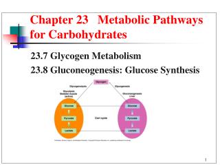
Three Steps Involved in Glucose Metabolism Glucose Anaerobic production of ATP. Occurs in the cytosol. However, not much ATP is produced by glycolysis, only two ATP molecules for each glucose molecule metabolized. Glycolysis ATP Pyruvate, NADH Found in all aerobic organisms, takes place in mitochondria of eucaryotes. Most of the coenzyme NADH is made here Through a series of redox reactions (NAD+ is reduced). Citric Acid Cycle NADH, FADH2 Found in eucaryotes, takes place in mitochondria. O2 is consumed by the electron transport chain. Most of the ATP is produced here, 28 ATP molecules for each glucose molecule metabolized. Oxidative Phosphorylation ATP
Energy is Invested at the Beginning of Glycolysis Two ATP molecules are used to make one molecule of FBP (G6P) (F6P) (PFK) (FBP)
Energy is Generated During Second Step (GPDH) Two ATP molecules produced for each of two glyceraldehyde-3- phosphate molecules, total of 4 ATP generated. Net ATP:
Glycolysis Can Be Oscillatory Sustained NADH oscillations in yeast, very simple (single cell) eucaryotes. Oscillations are in the presence of glucose and cyanide (which blocks electron transport, inhibiting oxidative phosphorylation). Dano et al., Nature, 402:320, 1999 Oscillations in three glycolytic intermediates in muscle extracts. Tornheim, JBC, 263:2619, 1988
What is the Mechanism for Glycolytic Oscillations? In muscle extracts the mechanism is known to be the allosteric enzyme Phosphofructokinase (PFK). The key feature of this enzyme is that its product FBP feeds back and stimulates the enzyme. The muscle form of this enzyme, PFK-M, dominates the PFK activity in b-cells.
Model Glycolytic Oscillations With moderate glucokinase activity With high glucokinase activity Bertram et al., Biophys. J., 87:3074, 2004 JGK is the glucokinase reaction rate JPFK is the PFK reaction rate JGPDH is the GPDH reaction rate
0.1 ratio 5 min Glycolytic Oscillations Occur Only for Moderate GK Rates 15 mM A model prediction is that it should be possible to turn on the GOs by simply increasing the glucose concentration. We have evidence for this from Ca2+ measurements in islets: 8 mM 8 mM Nunemaker et al., Biophys. J., 91:2082, 2006
Coenzymes are Produced by the Citric Acid Cycle Acetyl group has 2 carbons Oxaloacetate has 4 carbons Citrate has 6 carbons As the cycle progresses, first one carbon is lost and then another Cycle ends where it began, except that 4 NADH, one FADH2, and one GTP molecule have been made The coenzymes NADH and FADH2 are electron carriers that are used to transfer electrons between molecules. This transfer is key for powering oxidative phosphorylation
Anaplerosis and Cataplerosis Anaplerosis is a series of enzymatic reactions in which metabolic intermediates enter the citric acid cycle from the cytosol. Cataplerosis is the opposite, a process where intermediates leave the citric acid cycle and enter the cytosol. In muscle, anaplerosis is important for increasing citric acid throughput during periods of exercise. There is some evidence that anaplerosis is required for a glucose-induced rise in mitochondrial ATP production. Some amino acids (the building blocks of proteins) enter and leave the citric acid cycle through anaplerosis and cataplerosis.
Anaplerosis Involving Pyruvate Pyruvate pyruvate carboxylase
Anaplerosis Involving Amino Acids Leucine + Glutamate Glutamine GDH Histidine Proline Arginine
Anaplerosis Involving Amino Acids Valine Isoleucine Methionine
Anaplerosis Involving Amino Acids Phenylalanine Tyrosine
Anaplerosis Involving Amino Acids Aspartate Asparagine
Cataplerosis of Malate Phosphoenolpyruvate (PEP) Oxaloacetate Malate
Cataplerosis of Citrate Malonyl CoA Acetyl-CoA Oxaloacetate Fatty Acids
Subway Analogy • Citric Acid Cycle is like a subway system: • Acetyl-CoA is like people getting on at station A • NADH is like people getting off at station B • Intermediates are like the subway cars • Anaplerosis is like adding cars to the system • Cateplerosis is like removing cars to use for spare parts
The Malate/Aspartate Shuttle Some of the coenzyme NADH is made during glycolysis. How does this get into the mitochondria where it can power oxidative phosphorylation? 3 2 4 5 6 4 1 7 OAA=oxaloacetate MDH=malate dehydrogenase Asp=aspartate Glu=glutamate
Last Stage of Glucose Metabolism Produces the Most ATP Keeping score of ATP production: Glycolysis – 2 ATP for each glucose molecule Citric Acid cycle – No ATP produced Oxidative Phosphorylation – up to 34 ATP molecules Without mitochondria (and thus OP), complex life forms could not exist.
The Magnus-Keizer Model Published as a series of papers in the late 1990s. Describes oxidative Phosphorylation in b-cells. We have recently published a simpler model that uses curve fitting to reduce the complexity of the flux and reaction functions (Bertram et al., J. Theoret. Biol., 243:575, 2006). Mitochondrial Variables: NADH concentration ADP or ATP concentration (ADP+ATP=constant) Calcium concentration Inner membrane potential O2 consumption is also calculated
The NADH Equation NADH flux from citric acid cycle increases NADH concentration. NADH is oxidized when it supplies electrons to the electron transport chain, decreasing NADH concentration. JH,res Mitochondrial inner membrane Jo JDH
NADH Concentration Can Be Measured in Islets NADH autofluorescence is measured Bertram et al., Biophys. J., 92:1544, 2007
The ADP/ATP Equations ADP is phosphorylated to ATP by the F1-F0 ATP-synthase. This is due to the flux of protons down the concentration gradient from outside to inside of the mitochondrial inner membrane. The ATP made in this way is transported out, and ADP transported in, by the adenine nucleotide transporter. JANT JF1F0 ATP H+ ATP ADP
Cytosolic ATP Can Be Measured in Single b-cells ATP measured using adenovirally driven expression of recombinant firefly luciferase. Ainscow and Rutter, Diabetes, 51:S162, 2002
The Ca2+ Equation Calcium enters the mitochondria from the cytosol through calcium uniporters. Calcium is pumped out of the mitochondria into the cytosol via Na+/Ca2+ exchangers. Ca2+ JNaCa Juni Ca2+
The Inner Membrane Potential Equation This membrane potential is the driving force for ATP production by the F1F0 ATP synthase. If membrane potential is 0, then no ATP will be made. (Negative terms represent positive charge entering across the inner membrane) JH,leak JH,res JH,ATP JNaCa Juni JANT
Mitochondrial Inner Membrane Potential Can Be Measured in Islets Measured using the fluorescent dye rhodamine 123 (Rh 123) Kindmark et al., J. Biol. Chem., 276:34530, 2001
O2 Can Also Be Measured in Islets Measured using an oxygen electrode Kennedy et al., Diabetes, 51:S152, 2002