Download
1 / 55
550 likes | 637 Views
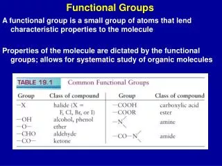
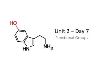
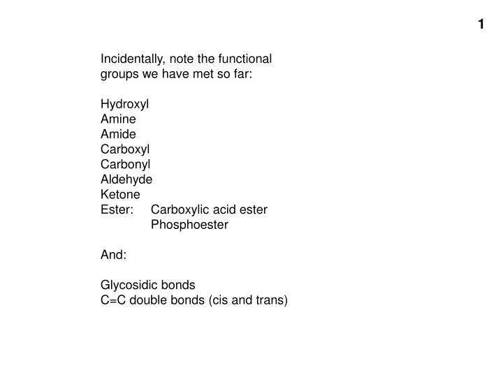
Incidentally, note the functional groups we have met so far: Hydroxyl Amine Amide Carboxyl Carbonyl Aldehyde Ketone Ester: Carboxylic acid ester Phosphoester And: Glycosidic bonds C=C double bonds (cis and trans). PROTEINS. Amino acids (the monomer of proteins).

E N D
Incidentally, note the functional groups we have met so far: Hydroxyl Amine Amide Carboxyl Carbonyl Aldehyde Ketone Ester: Carboxylic acid ester Phosphoester And: Glycosidic bonds C=C double bonds (cis and trans)
PROTEINS Amino acids (the monomer of proteins)
At pH 7, ,most amino acids are zwitterions (charged but electrically neutral)
+OH- ( -H+) +H+ 50-50 charged-uncharged at ~ pH2.5 (=the pK) 50-50 charged-uncharged at ~ pH9 (=the pK) Net charge
Numbering (lettering) amino acids ε-amino group ε δ γ β Alpha-carboxyl (attached to the α-carbon) Alpha-amino Alpha-carbon
Amino acids in 3 dimensions • Asymmetric carbon (4 different groups attached) • Stereoisomers • Rotate polarized light • Optical isomers • Non-superimposable • Mirror images • L and D forms From Purves text
Handout 3-3 (Without showing the R-groups) The backbone is monotonous It is the side chains that provide the variety
“Polypeptides” vs. “proteins” • Polypeptide = amino acids connected in a linear chain (polymer) • Protein = a polypeptide or several associated polypeptides (discussed later) • Often used synonymously • Peptide (as opposed to polypeptide) is smaller, even 2 AAs (dipeptide)
The backbone is monotonous (Without showing the R-groups) It is the side chains that provide the variety
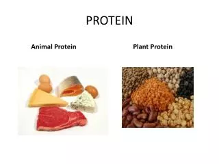
Proteins do most of the the jobs in the cell E.g., egg albumin, hemoglobin, keratin, estrogen receptor,immunoglobulins (antibodies), enzymes (e.g., beta-galactosidase) Each is a polymer or assemblage of polymers made up of amino acidsEach particular protein polymer (polypeptide) has a unique sequence of amino acidsEach molecule of a particular protein has the same sequence of amino acids. E.g., met-ala-leu-leu-arg-glu-leu-val- . . . . How is this sequence determined?
Primary (1o) Structure = the sequence of the amino acids in the polypeptide chain
asp ser Determining the sequence One way: enzyme: Carboxypeptidase: hydrolyzes the peptide bond , identify e.g., …. arg-leu-leu-val-gly-ala-gly-phe-trp-lys-glu-asp-ser …. arg-leu-leu-val-gly-ala-gly-phe-trp-lys-glu-asp …. arg-leu-leu-val-gly-ala-gly-phe-trp-lys-glu
AA mixture (-) (+)
Side view AAs applied at lower end
After stopping the paper chromatography and staining for the amino acids: “Rf” 0.82 0.69 0.45 0.27 0.11
Sub-peptides • Treatment of a polypeptide with trypsin • Trypsin is a proteolytic enzyme. • It catalyzes cleavage (hydrolysis) after lysine and arginine residues Polypeptide chain
The order of the subpeptides is unknown. The sequence is reconstructed by noting the overlap between differently produced subpeptides Trypsin (lys, arg) (1) Chymotrypsin (trp, tyr, phe) N C (2)
Hemoglobin protein Application to sickle cell disease (Ingram, 1960’s) Fingerprinting a protein: analysis of the sub-peptides (without breaking them down to their constituent amino acids) Sub-peptides No further digestion to amino acids; left as sub-peptides
Oligopeptides behave as a composite of their constituent amino acids H H H + H H H - H - Net charge = -1: moves toward the anode in paper electrophoreses Fairly hydrophobic (~5/6): expected to move moderately well in paper chromatography Nomenclature: ala-tyr-glu-pro-val-trp or AYEPVW or alanyl-tyrosyl-glutamyl-prolyl-valyl-tryptophan
The mixture of all sub-peptides formed Less negatively charged, More hydrophobic Hb In fingerprinting, these spots contain peptides, not amino acids trypsin Negatively charged ---valine--- (sickle) Positively charged More hydrophobic ---glutamate--- (normal) More hydrophilic Negatively charged Positively charged Negatively charged Positively charged
Every different polypeptide has a different primary structure (sequence). Every polypeptide will have different arrangement of spots after fingerprinting.
3-dimensional structure of proteins One given purified polypeptide • Molecule #1: N-met-leu-ala-asp-val-val-lys-.... • Molecule #2: N-met-leu-ala-asp-val-val-lys-... • Molecule #3: N-met-leu-ala-asp-val-val-lys-... • Molecule #4: N-met-leu-ala-asp-val-val-lys-... etc .
3-D structure of polypeptides Arrangement 3 at one momentin time? Arrangement 1 at one momentin time? Arrangement 2 at one momentin time? Arrangement 3 at one momentin time? [Rope models here]
Primary structure itself results in some folding constraints: See bottom of handout 3-3
4 atoms in one plane 6 atoms in one plane
Secondary structure: the alpha helix Amino acids shown simplified, without side chains and H’s.
Alpha helix depictions C = grays N = blue O = red Poly alanine Side chains = -CH3 (lighter gray) H’s not shown
H-bond AA residue Secondary structure:
Beta-sheets Anti-parallel Parallel
secondary structure (my definition): structure produced by regular repeated interactions betweenatoms of the backbone.
Neither Beta-sheets Alpha-helices Tertiary structure: The overall 3-D structure of a polypeptide. These “ribbon” depictions do not show the side chains, only the backbone