Download
1 / 48
480 likes | 499 Views
Explore how hormones maintain homeostasis and regulate metabolism. Learn about glands, signal transmission, feedback mechanisms, and the crucial role of the hypothalamus and pituitary gland. Discover the functions of the hypophysis, hormone secretion regulation, and more.

E N D
The endocrine system Department of Anatomy, Histology and Developmental Biology Faculty of Medicine, Semmelweis University 2019. Dr. Csáki Ágnes
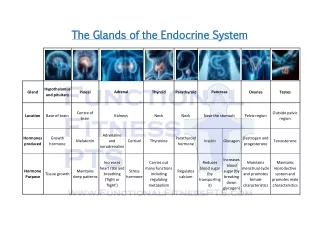
The glands of the endocrine system secrete hormones into the bloodstream to maintain homeostasis and regulate metabolism. Hormone: -minute amount of chemical substances (amines, steroids, peptides, proteins ) coordinate metabolic processes in cells and organs by activating enzymes (although each hormone is distributed by the blood , only particular cells with a specific receptor for it react to each individual hormone) (membrane or cytoplasmic receptors) -hormones are also produced by neurons of CNS and PNS: neurohormones -are vital substances [hyper or hyposecretion of a hormone leads to typical disease patterns]
MECHANISM OF SIGNAL TRANSMISSION endocrine neuroendocrine neuron cell capillary neurohormon hormon parakrin cell autokrin
Tissues and individual cells secreting hormons Classic endocrine organs Central and autonomic nervous System Parts of the diencephalon (e. g., hypothalamus) System of gastrointestinal cells Atria of the heart Atrial natriuretic peptide Kidneys Liver Immune organs (thymus) Tissue hormones C cells Ovary placenta
Types of glands endocrine exocrine
Regulation of Hormone Secretion The hypothalamus and the pituitary gland are the command and control centers, directing hormones to other glands and throughout the body The stability of the blood level of a hormone is the result of a complicated regulating mechanism: „feedback„ mechanism: It is a process in which the level of one substance influences the level of another substance. A mechanism or a signal that tends to initiate (or accelerate) or to inhibit (or slow down) a process. hormon level falls - more of it is released if it rises - less hormone is produced Negative feedback:
Other brain areas (limbic system, sensory organs..) hypothalamus hypophysis glands cells FEED-BACK MECHANISM Hyper or hyposecretion of a hormone leads to typical disease patterns ultrashort feedback (neural) hypophyseotroph neurons ultrashort feedback (humoral) anterior lobe troph hormones short feedback long feedback Béla Halász,in: Handbook of Clinical Endocrinology and Metabolic Diseases
The hypothalamus and the pituitary gland are part of the diencephalon region of the brain. The hypothalamus connects the nervous system to the endocrine system. It receives and processes signals from other brain regions and translates them into hormones. These hormones flow to the pituitary gland, which is connected to the hypothalamus Some hypothalamic hormones are stored in the pituitary for later release; others spur it to secrete its own hormones. Master of endocrine glands: Hypothalamus („real boss”) and Hypophysis („master gland”)
FUNCTIONS OF THE HYPOTHALAMUS Regulatesthehypophysis Servesas an endocrineorgan: oxytocyn, vasopressinsecretion Otherfunctions: Integrator and modulator of autonomicactivity (byzones and regions) - sympathetic and parasympatheticcenters (stress) - controlthe body temperature - regulation of food and waterintake etc. Circadianrythmthroughbiologicalclock (jet leg) Sexualbehavior and reproduction
HYPOTHALAMUS MEDIAN SAGITTAL SECTION OF THE BRAIN Position of hypothalamus CEREBRAL HEMISPHERE THALAMUS III.v. CEREBELLUM BRAIN STEM STALK- pituitary gland / hypophysis
HYPOTHALAMUS INFERIOR VIEW Visual system: optic chiasm hypophyseal stalk optic tract Hypophyseal tumors!! These benign tumors do not spread outside the skull. They usually remain confined to the sella turcica (the tiny space in the skull that the pituitary gland sits in). There is very little room for tumors to grow in this part of the skull. Therefore, if the tumor becomes larger than about a centimeter (about half an inch) across, it may grow upward, where it can compress and damage nearby parts of the brain and the nerves that arise from it. This can lead to symptoms such as vision changes or headaches.
Median sagittal MRI section with the hypophysis hypothalamus Air-filled sphenoidalis sinus (sphenoid bone) nasal cavity tongue cerebellum brain stem
SELLA TURCICA pituitary gland Operation of the hypophysis through the nasal cavity and sphenoidal sinus X-ray film
Hypophysis, pituitary gland Known as the "Master Gland", this part of the brain consists of two parts called adeno hypophysis and neurohypophysis. hypothalamus 1.Neurohypophysis (posterior part) Nervous tissue Optic chiasm pituitary stalk Only storage of hormones produced by hypothalamus 2.Adenohypophysis (Anterior and intermediate part) Glandular tissue Producing of hormones sella turcica / fossa hypophysealis/ bone
Two groups of the cells in the hypothalamus: magnocellular and parvocellular nuclei, act on different parts of the hypophysis
„magnocellular” nuclei (groups of big neurons): secretion of oxytocyn and antidiuretic hormon (ADH) /vasopressin / axonal transport to the hypophysis and stored in the posterior lobe and released to the capillaries
FUNCTION OF POSTERIOR PITUITARY HORMONES OXYTOCIN Contraction of uterinal smooth muscle (delivery) Contractionof the mammary gland smooth muscle (milk ejection) VASOPRESSIN (antidiuretic hormone) Vasoconstrictor, enhances the blood pressure Water absorption in the collective tubules of kidney
pituitary stalk „parvocellular”nuclei (groups of small neurons ) Releasing or inhibiting hormones RELEASING HORMONES (factors) LHRH (luteinizing hormone-releasing hormone) TRH (thyreotrop hormone-releasing hormone) CRH (corticotrop hormone-releasing hormone) GRH (growth hormone-releasing hormone) INHIBITING HORMONES (factors) Somatostatin (growth hormone-inhibiting hormone) Dopamine (prolactin-inhibiting hormone) Parvocellular PORTAL CIRCULATION OF THE ANTERIOR PITUITARY portal vessels Hormones of the adenohypophysis: LH: luteinizing hormone FSH: follicle stimulating hormon ACTH: adrenocorticotropin h TSH: thyroid stimulating h GH: somatotrpic h MSH: melanocyte stimulating h PRL: prolactin (mammotropic) anterior pituitary (adenohypophysis- gland) Béla Halász, in: Handbook of Clinical Endocrinology and Metabolic Diseases
Hystology of the hypophysis Anterior and intermediate lobe: Real gland ADENOHYPOPHYSIS Chromophil cells: well stained Basophil and acidophil cells produce hormones Chromophob cells poorly stained unknown function Posterior lobe : NEURO- HYPOPHYSIS Axons, glial cells and capillaries Capillaries, sinusoids
HORMONES OF THE ANTERIOR LOBE GH (growth hormone) LH (luteinizing hormone) - acidophil cells FSH (follicle stimulating hormone) TSH (thyroid stimulating hormone) ACTH (adrenocorticotrop hormone) LTH (luteotrop hormone, prolactin) - basophil cells Interleukins, growth factors (folliculostellate cells)
(Anterior lobe) Growth hormon (GH) Acidophil Cell growth hormon induces growth throught insuline like factor produced by hepatocytes. IGF-1 stimulates the growth of long bones by stimulating the hypertrophy of epiphyseal plates
Inadequate production of somatotropin (GH) maybe • Hypo- or Hyperproduction Hypo: Pituitary dwarfism dwarf (neat, proportional) normal size
Hyperproduction of somatostatin (GH) GIGANTISM ACROMEGALY in adults, after full growth has ceased (Ill-proportioned, Enlargement of the bones in the jaws and in the front of the skull are typically the most apparent bony changes. Acromegaly may also cause thickening of the soft tissues of the body, including the heart, lips, and tongue. normal giant normal high level from childhood
(Anterior lobe) Prolactin Acidophil cells
(Anterior lobe) FSH(folliculus stimulating hormon) LH (luteinizing hormon) Basophil cells Stimulates development of Graafin follicles (= a mature follicle in the ovary prior to ovulation) Brings about ovulation and maintains the corpus luteum.
(Anterior lobe) TSH (thyroid stimulating hormon) Basophil cells Stimulates the thyroid to release T3 and T4.
(Anterior lobe) ACTH Basophil cell Stimulates the adrenal cortex to produce:Corticosteriods: mineral corticoids glucocorticoids cortisol (natural anti-inflammatory !) androgens, e.g. aldosterone
1. Pituitary-dependent endocrine organs Thyroid gland Adrenal gland (Ovary or testis) 2. Pituitary-independent endocrine organs and cells Pineal body Parathyroid gland Islets of Langerhans (Placenta) (Enteroendocrine cells – cells in the gastrointestinal tract) Central endocrine organ: pituitary gland, hypothalamus Peripheral endocrine organs
1. Pituitary-dependent endocrine organs Thyroid gland At the trachea (in its capsule there are four parathyroid glands !) Hypothalamic TRH Hypophyseal TSH Trijodthyronin(T3), thyroxin(T4) (C cells produce calcitonin - independent)
Histology of the thyroid gland colloid in follicles T4,T3 immunostaining for calcitonin in C-cells Calcitonin lowers the calcium level of the blood and promotes bone formation hematoxylin-eosin staining
absorption by endocytosis colloid follicular epithelium lysosome colloid (thyroglobulin) Thyroxin (T4) Triiodothyronin (T3) RER capillary amino acids + iodine Mechanism of Thyroid hormone production SYNTHESIS iodine Functional phases of the follicular cells: -secretion is being formed -during storage -during the phase of secretion
Hypo- and hyperfunction of the gland: BASEDOW-DISEASE (hyperthyreosis) CRETENISM (hypothyreosis from childhood) bigger and become visible as a “goiter”, also called “struma.” Mentally retarded It is particularly important for children and babies to have enough thyroid hormones (jodine!!!), because a lack of these hormones at an early stage of life can have severe effects on physical and emotional development
Adrenal gland Adrenal gland cortex medulla kidney Section of the gland vein cortex medulla
Adrenal gland 3 layers of the cortex zonaglomerulosa zonafasciculata cortex zonareticularis medulla
Zona reticularis Zona glomerulosa Zona fasciculata Glükocorticoids-cortisol sexual steroids mineralocorticoids MEDULLA: neuronal cells produce EPINEPHRIN, NOREPINEPHRIN sympathetic system!! involved in defense reaction to stress (hypothalamic-pituitary-adrenal (HPA) axis)
Cushing's syndrome is a collection of signs and symptoms due to prolonged exposure to cortisol. Signs and symptoms may include: high blood pressure, abdominal obesity but with thin arms and legs, reddish stretch marks, a round red face, a fat hump between the shoulders, weak muscles, weak bones, acne, and fragile skin that heals poorly. Women may have more hair and irregular menstruation. Occasionally there may be changes in mood, headaches, and a chronic feeling of tiredness. Cushing syndroma Overproduction of zona fasciculata
2. Pituitary-independent endocrine organs HISTOLOGY brain sand Pinealocytes Glial cells capillaries PINEAL BODY or epiphysis Part of the thalamus calcified structures Melatonin: produced from serotonin, restricts the release of gonadotropins, restrains development of gonads, subfunctional pineal body –pubertas precox: early sexual maturation Regulates the day and night rythm,
Parathyroid glands Oxyphil cell: unknown function Chief cells Fat cell (Thyroid gland) Chief cells produce parathormone: controls the concentration of calcium and phosphate ions of blood: elevates the blood Ca level: antagonist of the Calcitonin!!
In the pancreas – Langerhans islets spleen kidney duodenum aorta Inferior vena cava
LANGERHANS-islets in the pancreas (about 1 million) • A-cells: GLUCAGON • B-cells: INSULIN • regulate glucose levels in the blood • D-cells: SOMATOSTATIN • P-cells: PANCREAS-POLYPEPTID • Exocrin pancreas • Tripsine • Kimotripsine • Carboxypeptidase • Pancreas amilase • Pancreas lipase • DNA-se, RNA-se
Thank for your attention! Szentágothai János, Réthelyi Miklós: Funkcionális anatómia Kiss Ferenc, Szentágothai János: Az ember anatómiájának atlasza Johannes W. Rohen, Chihiro Yokochi, Elke Lütjen-Drecoll: Color atlas of anatomy Röhlich Pál: Szövettan Leövey János: Handbook of clinical endocrinology and metabolic diseases
T3 and T4 increase the basal metabolic rate. All body cells then work harder and therefore need more energy. This means: Body temperature rises The heart beat becomes stronger and the pulse faster Food is used up more quickly because energy stored in the liver and muscles is broken down Brain maturation is promoted (in children) Growth is promoted (in children) Activation of the nervous system leads to higher levels of attention and quicker reflexes
With an overactive thyroid, also called hyperthyroidism, it produces too many hormones. This speeds up energy metabolism, and the following symptoms can occur: Hot flashes, sweating Trembling Weight loss Diarrhea Hair loss Nervousness, hyperactivity Emotional instability and irritability or fatigue Insomnia and restlessness Potency problems
Thyroid underactivityhypothyroidism in adults often develops gradually, which is why people might not notice symptoms for a while. Possible symptoms can include: General loss of energy and power Slowed metabolism Being overweight Tiredness Difficulties concentrating or mental slowness Constipation Sensitivity to cold Slow pulse Waxy skin thickening and swelling (myxedema) Dry skin Deep, hoarse voice Brittle, dry hair Loss of sexual desire or potency problems Sometimes even depression