Download
1 / 53
540 likes | 702 Views
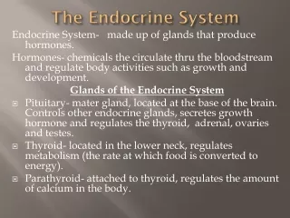
THE ENDOCRINE SYSTEM. Kristina C. Erasmo, M.D. ENDOCRINE SYSTEM. System of glands that involve the release of extracellular signaling molecules known as hormones Do not possess any excretory ducts Integration and control system of the body. Hormone.

E N D
THE ENDOCRINE SYSTEM Kristina C. Erasmo, M.D.
ENDOCRINE SYSTEM • System of glands that involve the release of extracellular signaling molecules known as hormones • Do not possess any excretory ducts • Integration and control system of the body
Hormone • Substance that is carried by the blood to target organs/tissues whose cells have the appropriate receptors for the substance • Function as “chemical signals”
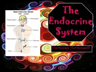
ENDOCRINE SYSTEM • Hypothalamus • Pituitary gland • Pineal gland • Thyroid gland • Parathyroid gland • Adrenal gland • Pancreas
HYPOTHALAMUS • At base of brain, behind the optic chiasm • Forms the floor and part of the wall of the 3rd ventricle • Part of the diencephalon
HYPOTHALAMUS • Functions: • Control numerous bodily functions • Thirst, hunger, satiety • Temperature • Sexual behavior • Circadian rhythms • Produces several hormones • Oxytocin • Antidiuretic hormone • Neurohormones
HYPOTHALAMUS • The cell bodies of the neurons form numerous aggregations (nuclei) • Supraoptic nucleus • Above and lateral to optic chiasm • Paraventricular nucleus • In the lateral wall of 3rd ventricle
HYPOTHALAMUS • Oxytocin • Stimulates contractions of the uterus and ejection of milk • Vasopressin (antidiuretic hormone/ADH) • Regulates water retention (kidneys) • ↑ urine osmolarity (↑ concentration), ↓ urine excretion • Neurohormones (hypophysiotropic hormones) – secretory neurons
PITUITARY GLAND • Ovoid body attached to inferior surface of hypothalamus • Lodged in the hypophysealfossa of the sellaturcica of the sphenoid bone
PITUITARY GLAND: PARTS • Neurohypophysis (posterior pituitary) • From neural ectoderm • Adenohypophysis (anterior pituitary) • From oral ectoderm
Neurohypophysis • Greater part is formed by the axons of the secretory neurons whose cell bodies are in the SVN and PVN of hypothalamus • Proximal portions of these axons form hypothalamo-hypophyseal tract • Distal portions form bulk of the posterior lobe of pituitary
Neurohypophysis • Not a gland • Stores and secretes oxytocin and ADH (“posterior pituitary hormones”) • 2 parts: • Pituitary stalk (infundibulum, infundibular stem, infundibular stalk, hypophyseal stalk) • Attached to the hypothalamus • Pars nervosa (posterior lobe, infundibular process) • Expanded inferior continuation of the pituitary stalk
Adenohypophysis • Produces “anterior pituitary hormones” • Somatotropin, prolactin, thyrotropin, corticotropin, FSH, LH • 3 parts: • Pars distalis(anterior lobe) • Pars tuberalis(pars infundibularis) • Pars intermedia(intermediate lobe)
Pars Tuberalis • Forms a sleeve around the pituitary stalk of neurohypophysis • Where vessels of hypophyseoportal system and arteries that supply anterior and posterior lobe run through
Pars Intermedia • Thin, poorly developed region of adenohypophysis • Contain secretory granules • Melanocyte-stimulating hormone (MSH) • β-endorphin
Pars Distalis • Comprises 70% of pituitary gland • Nearly all hormones produced by pituitary gland come from this lobe (“anterior pituitary hormones”)
Pars Distalis • Consists of anastomosing and irregularly arranged cords and clusters of secretory epithelial cells that surround fenestrated capillaries
Pars Distalis • 2 types of secretory cells: • Chromophils • Acidophils (alpha cells) • Basophils (beta cells) • Chromophobes
Chromophobes • 65 % of epithelial secretory cells in pars distalis • Consists of follicular cells and undifferentiated stem cells
Chromophils • 35 % of epithelial secretory cells of pars distalis • Acidophils • Somatotrophs • Mammotrophs • Basophils • Thyrotrophs • Corticotrophs • Gonadotrophs
Hypophysiotropic Hormones • Hypothalamic control over anterior gland of pituitary gland • Corticotropin-releasing hormone (CRH) • Thyrotropin-releasing hormone (TRH) • Growth hormone-releasing hormone (GRH) • Growth hormone-inhibiting hormone (GIH, somatostatin) • Gonadotropin-releasing hormone (GnRH) • Prolacting-inhibiting hormone (PIH)
PINEAL GLAND • Small, conical structure • Base is attached to posterior portion of the roof of the 3rd ventricle of the brain by two short stalks • Well-developed in children • Starts to involute at puberty
PINEAL GLAND • Enveloped by thin CT capsule derived from the pia mater • CT septa arise from the capsule and divide the organ into irregular lobules • Lobules contain cells arranged in short cords or clusters surrounded by fenestrated capillaries • CT septa contain blood vessels and unmyelinated nerve fibers
PINEAL GLAND • Cells: • Pinealocytes (95 %) • Modified neurons • Produce melatonin • Glial cells (Interstitial cells) • Brain sand (Psammoma bodies) • Calcified bodies with a concentric lamellar structure • Increase in number with age
THYROID GLAND • Largest endocrine gland • Unpaired gland that lies on the anterior aspect of the neck • Left and right lateral lobes • Isthmus – horizontal bridge of glandular tissue which connects both lobes
THYROID GLAND • Enclosed by two capsules • CT septa divide the organ into lobules • CT infiltrated with lymphocytes and some lymphoid nodules • Each lobule contains follicles
THYROID GLAND • Follicles • Irregular spherical, cystic structures • Lined by simple cuboidal epithelium • Cavity contains colloid (homogenous, gel-like material)
THYROID GLAND • Epithelial cells: • Follicular cells (principal cells) • Majority • Synthesizethyroglobulin • Parafollicular cells (C or clear cells) • 0.1 % • Synthesize calcitonin– inhibits bone resorption (directly suppressing osteoclasts), thus lowering blood calcium levels
PARATHYROID GLAND • Two pairs (superior and inferior), yellowish-brown, tiny ovoid bodies • Attached to the posterior surface of the lateral lobes of the thyroid gland
PARATHYROID GLAND • Chief cells (principal cells) • Majority • Secrete parathyroid hormone (PTH) • Oxyphil cells (acidophil cells) • Non-secretory • Unknown function
ADRENAL GLAND • Paired, flat, pyramidal organs • Rest on the upper pole of their correspondin kidneys
ADRENAL GLAND • Enveloped by thick CT capsule • Cortex • Outer layer • Completely surrounds medulla • Mesodermal origin • Medulla • Inner layer • Ectodermal origin
Adrenal Cortex • 80 – 90 % of the adrenal gland • Produce adrenocortical hormones (steroid hormones) • Zonaglomerulosa– aldosterone • Zonafasciculata– cortisol • Zonareticularis– androgens (DHEA and androstenedione)
Adrenal Medulla • Comprises 10-20 % of adrenal gland • Central part: medullary veins that drain the entire gland • Consists of parenchymal cells arranged in groups or cords surrounded by sinusoids and richly supplied with nerves (associated with pre-ganglionic sympathetic neurons)
Adrenal Medulla • Chromaffin cells: secrete cathecolamines • Epinephrine (adrenaline) • Norepinephrine (noradrenaline) • Dopamine
PANCREAS • Islets of Langerhans • Small aggregations of pale-staining cells scattered among the dark-staining cells of the exocrine portion of the pancreas • Each islet surrounded by thin layer of fine reticular fibers • Provided by rich supply of capillaries • Millions but only 2 % of volume of pancreas