Download
1 / 42
530 likes | 685 Views
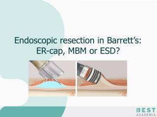
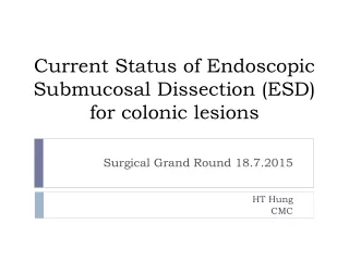
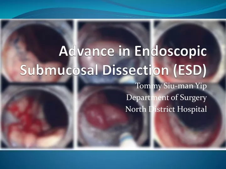
Advance in Endoscopic Submucosal Dissection (ESD). Tommy Siu-man Yip Department of Surgery North District Hospital. Background. Endoscopic submucosal dissection (ESD) first described in 1988 to treat early gastric cancer; now expanded to locations throughout GI tract

E N D
Advance in Endoscopic Submucosal Dissection (ESD) Tommy Siu-man Yip Department of Surgery North District Hospital
Background • Endoscopic submucosal dissection (ESD) first described in 1988 to treat early gastric cancer; now expanded to locations throughout GI tract • Enables en bloc removal of larger and potentially deeper lesions with a curative intent than can be accomplished with EMR
ESD steps during resection • 1) Marking of perimeter of lesion with cautery • 2) Injection of lifting agent into submucosa around the perimeter of lesion • 3) Incision of mucosa and circumferential cut around lesion with an eletrosurgical knife • 4) Injection of submucosa beneath lesion and dissection until complete resection of specimen • 5) Hemostasis and vessel coagulation
Knives A: ITKnife. B: ITKnife2. C: ITKnife nano. D: HookKnife. E: TTKnife. F: DualKnife G: FlexKnife. H: HybridKnife I type. I: HybridKnife T type
Hemostatic forceps Coagrasper hemostatic forceps: Colonic forceps (left) and gastric forceps (right)
Cap • A transplant distal attachment uniformally applied to the tip of endoscope • Maintain visualization by keeping the resected flap of mucosa off the endoscope lens • Many caps feature drainage holes to allow egress of water and blood
Dyes • Better characterize the surface of lesion • Clearer demarcation of borders • Better recognition of tissue planes during dissection by coloring the injectate for submucosal lifting • E.g Indigo carmine, methylene blue, Lugol’s iodine
Injection agents Drug Des Devel Ther. 2008; 2: 131–138.
Endoscopes • Influenced by a number of considerations • Auxillary water channel with flushing pump for water-jet effect to maintain visualization • Two instrument channels • Allow dual instrument use • Choice of channel for optimal working angle • Extra channel for suctioning • ? High definition ? High magnification • ? Multi-bending endoscope
Gas insufflation • Carbon dioxide (CO2) • Absorbed across the intestines rapidly • Less prolonged luminal distension and less patient discomfort • Reduce the likelihood of tension pneumoperitoneum in case of perforation
Sedation • From moderate sedation (eg. midazolam), deep sedation (eg. propofol) or general anesthesia (GA) • GA for upper GI lesions • Potential for reflux and aspiration of secretion or blood • Moderate to deep sedation for colorectal ESD • Conscious sedation may facilitate changes in posture to use gravity as counter-traction
Complications of ESD • Bleeding • Perforation • Stricture
Bleeding • Coagulation by ESD knife / hemostatic forceps • Delayed bleeding more common in gastric ESD (4.5%, up to 15.6%) • PPI +/- mucosal protective agent • Urgent surgery
Perforation • 2.3-10% in oesophageal ESD 4.5% for gastric ESD 4.8% in colorectal ESD • Most amendable by clip closure • Gastric ESD • ≤1cm defect: Primary clip closure • Larger defect: “omental patch” method • NG tube, TPN, antibiotics • Urgent surgery
Stricture • 12-17% after oesophageal ESD • Highest risk if resection >75% circumference of oesophagus • Management • Prophylactic serial dilatation • Intralesional / topical steroid application • Prophylactic placement of SEMS
Advance in ESD • Problems to be tackled • Dissection • Traction • Ease of dissection of submucosal plane • Prevention of Complications
Advance in ESD • Problems to be tackled • Dissection • Traction • Ease of dissection of submucosal plane • Prevention of Complications
Anubiscope – An endoscopic surgical platform with two working channels, for surgical instruments with articulated tip allowing four degrees of freedom Master and slave transluminal endoscopic robot (MASTER)
Advance in ESD • Problems to be tackled • Dissection • Traction • Ease of dissection of submucosal plane • Prevention of Complications
Chemical dissection • Mesna (sodium 2-mercaptoethanesulfonate) • Thiol compound • Dissolve disulfide bonds in connective tissue between anatomical planes • Evaluated in a double-blind, randomized, placebo-controlled trial of 101 patients undergoing gastric ESD • Submucosal dissection time was 18.6 minutes in the mesna group and 24.6 minutes in the placebo group (P=0.13) • However, multivariate regression analysis found use of mesna to be highly correlated with submucosal dissection time Gastrointest Endosc 2014;79:756-64.
Cook Medical’s submucosal lifting gel • consist of a proprietary combination of known biocompatible components • safe and effective for a durable submucosal cushion without resulting in tissue damage in animal models
Advance in ESD • Problems to be tackled • Dissection • Traction • Ease of dissection of submucosal plane • Prevention of Complications
Endoscopic suturing of ESD defects • Overstitch endoscopic suturing device • Evaluated in a retrospective, single-center study of 12 patients (4 lesions in stomach, 8 lesions in colon; mean lesion size, 42.5 ± 14.8 mm) over a period of 8 months in 2012-13 • Technically feasible and fast (mean closure time, 10.0 ± 5.8 minutes per patient) • Patients discharged home on the same day after the procedure • No immediate or delayed adverse events observed
Endoscopic tissue shielding method • Polyglycolic acid (PGA) sheet & fibrin glue • Evaluated in a prospective, single-arm, pilot study in 10 patients with colorectal tumours in 2012 in a single tertiary centre in Japan • Mean tumor size 39.7 ± 15.2mm • Mean procedure time 18.7 ± 15.9 minutes • No procedure-related adverse events occurred • Upon colonoscopy 9 to 12 days after ESD, the PGA sheets were still fixed on the whole defect in 8 patients Gastrointest Endosc 2014;79:151-5.
Conclusion • ESD is an effective treatment modality for premalignant and early-stage malignant lesions of stomach, oesophagus and colorectum • ESD is technically demanding and has a higher rate of adverse events than most endoscopic procedures • Additional refinements in the technique, instruments, devices and training would be necessary to further reduce the higher risk of complications and longer procedure times
General indications for ESD • Oesophageal ESD: • Endoscopic signs of early lesions or echo endoscopic examination confirming tumor limited to the mucosa or up to the superficial submucosa (Sm1) • Histological confirmation of squamous cell carcinoma or high grade intraepithelial neoplasia restricted to the mucosa (M1 and M2). • Lesions with M3 or Sm1 invasion without lymphatic and vascular involvement, no larger superficial size (< 2.5 mm) • No signs of LN metastases
Colorectal ESD: • ≥2cm lesions in which en bloc resection by snare EMR difficult • LST-NG type particularly pseudo-depressed lesion • Kudo V pit pattern • Cancer with submucosal infiltration • Mucosal lesion with fibrosis • Local residual early cancer after endoscopic resection • Sporadic localized tumours in chronic inflammation such as UC