Download
1 / 1
10 likes | 120 Views
The importance and methods of dispersing fillers into epoxy resin M Reading * , Z. Xu, A S Vaughan and P L Lewin University of Southampton, Southampton, UK. Results. Introduction.

E N D
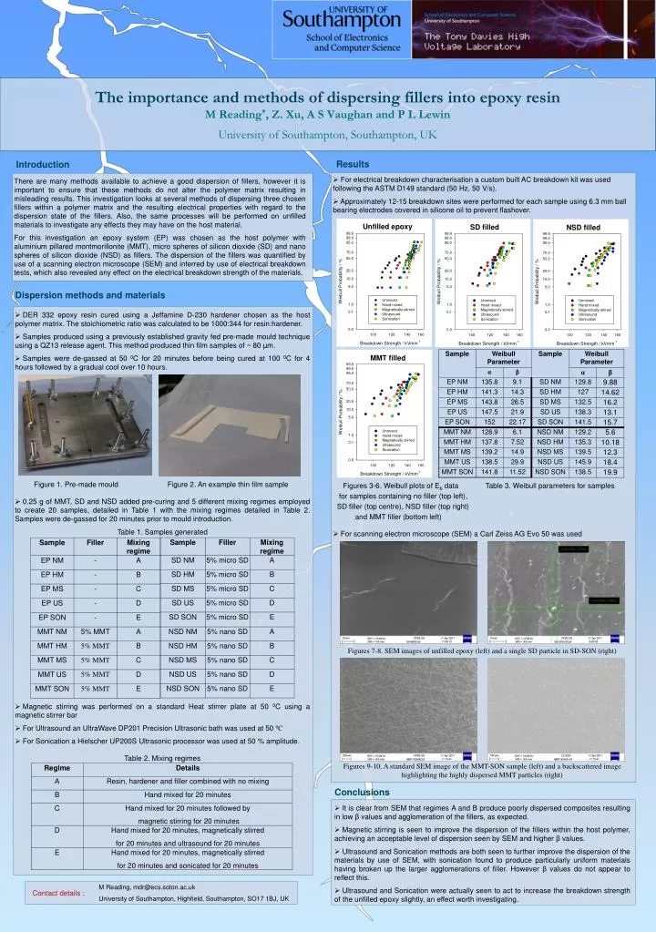
The importance and methods of dispersing fillers into epoxy resin M Reading*, Z. Xu, A S Vaughan and P L LewinUniversity of Southampton, Southampton, UK Results Introduction • For electrical breakdown characterisation a custom built AC breakdown kit was used following the ASTM D149 standard (50 Hz, 50 V/s). • Approximately 12-15 breakdown sites were performed for each sample using 6.3 mm ball bearing electrodes covered in silicone oil to prevent flashover. Figures 3-6. Weibull plots of Eb data Table 3. Weibull parameters for samples for samples containing no filler (top left), SD filler (top centre), NSD filler (top right) and MMT filler (bottom left) • For scanning electron microscope (SEM) a Carl Zeiss AG Evo 50 was used There are many methods available to achieve a good dispersion of fillers, however it is important to ensure that these methods do not alter the polymer matrix resulting in misleading results. This investigation looks at several methods of dispersing three chosen fillers within a polymer matrix and the resulting electrical properties with regard to the dispersion state of the fillers. Also, the same processes will be performed on unfilled materials to investigate any effects they may have on the host material. For this investigation an epoxy system (EP) was chosen as the host polymer with aluminium pillared montmorillonite (MMT), micro spheres of silicon dioxide (SD) and nano spheres of silicon dioxide (NSD) as fillers. The dispersion of the fillers was quantified by use of a scanning electron microscope (SEM) and inferred by use of electrical breakdown tests, which also revealed any effect on the electrical breakdown strength of the materials. Unfilled epoxy SD filled NSD filled Dispersion methods and materials • DER 332 epoxy resin cured using a Jeffamine D-230 hardener chosen as the host polymer matrix. The stoichiometric ratio was calculated to be 1000:344 for resin:hardener. • Samples produced using a previously established gravity fed pre-made mould technique using a QZ13 release agent. This method produced thin film samples of ~ 80 µm. • Samples were de-gassed at 50 0C for 20 minutes before being cured at 100 0C for 4 hours followed by a gradual cool over 10 hours. Figure 1. Pre-made mould Figure 2. An example thin film sample • 0.25 g of MMT, SD and NSD added pre-curing and 5 different mixing regimes employed to create 20 samples, detailed in Table 1 with the mixing regimes detailed in Table 2. Samples were de-gassed for 20 minutes prior to mould introduction. Table 1. Samples generated • Magnetic stirring was performed on a standard Heat stirrer plate at 50 0C using a magnetic stirrer bar • For Ultrasound an UltraWave DP201 Precision Ultrasonic bath was used at 50 0C • For Sonication a Hielscher UP200S Ultrasonic processor was used at 50 % amplitude. Table 2. Mixing regimes MMT filled Figures 7-8. SEM images of unfilled epoxy (left) and a single SD particle in SD-SON (right) Figures 9-10. A standard SEM image of the MMT-SON sample (left) and a backscattered image highlighting the highly dispersed MMT particles (right) Conclusions • It is clear from SEM that regimes A and B produce poorly dispersed composites resulting in low β values and agglomeration of the fillers, as expected. • Magnetic stirring is seen to improve the dispersion of the fillers within the host polymer, achieving an acceptable level of dispersion seen by SEM and higher β values. • Ultrasound and Sonication methods are both seen to further improve the dispersion of the materials by use of SEM, with sonication found to produce particularly uniform materials having broken up the larger agglomerations of filler. However β values do not appear to reflect this. • Ultrasound and Sonication were actually seen to act to increase the breakdown strength of the unfilled epoxy slightly, an effect worth investigating. M Reading, mdr@ecs.soton.ac.uk University of Southampton, Highfield, Southampton, SO17 1BJ, UK Contact details :