Download
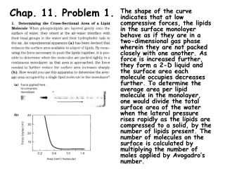
1 / 31
320 likes | 534 Views
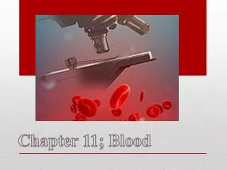
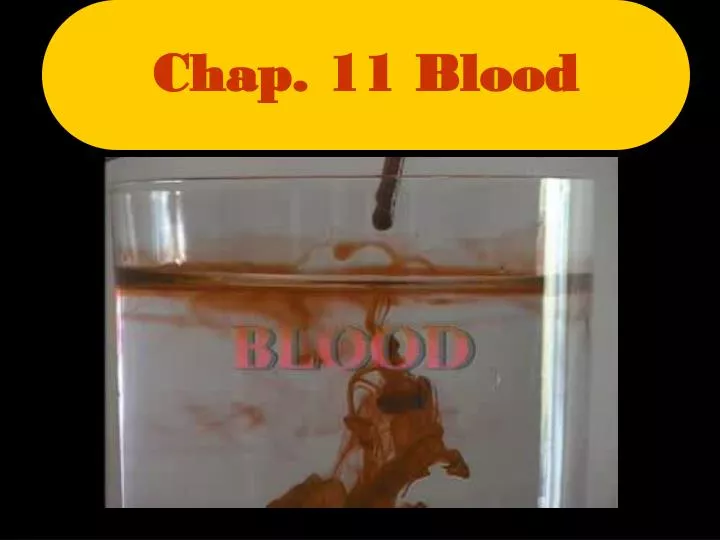
Chap. 11 Blood. 11-1 Functions of Blood. Transport of dissolved substances – gases, nutrients, hormones, metabolic wastes Regulation of pH and ions Restriction of fluid loss at injury site – blood clotting Defense against toxins and pathogens – white blood cells and antibodies

E N D
11-1 Functions of Blood • Transport of dissolved substances – gases, nutrients, hormones, metabolic wastes • Regulation of pH and ions • Restriction of fluid loss at injury site – blood clotting • Defense against toxins and pathogens – white blood cells and antibodies • Stabilization of body temp
11-1 Composition of Blood • Blood is the only fluid tissue in the body • Whole blood consists of living blood cells, called formed elements (37-54%), suspended in a nonliving fluid matrix called plasma (46-63%) • When spun in a centrifuge, a process known as fractionation, the erythrocytes(red blood cells), sink to the bottom. Their main function is oxygen transport. • Superior to this is the buffy coat, containing leukocytes (white blood cells) and platelets, which prevent infection • Superior to this is the pale yellow plasma. Erythrocytes
11-1 Physical Characteristics and Volume • Blood is a sticky, opaque fluid with a metallic taste • Oxygen rich blood has a scarlet color, while oxygen poor blood has a dull red color • Blood is heavier than water, because of the formed elements • High viscosity • Blood has a slightly alkaline pH of between 7.35 and 7.45 • Typical blood temperature is about 100.4oF (38oC) • Blood volume (liters) makes up about 7% of body weight • Adult male = 5-6 liters • Adult females = 4-5 liters
11-2 Plasma • Plasma is the liquid part of the blood • It is straw colored and is about 92% water • Plasma consists of over 100 dissolved substances such as nutrients, salts, hormones, and plasma proteins (most abundant) • Composition similar to interstitial fluid • Plasma helps to distribute heat evenly throughout the body The pale yellow substance is plasma
11-2 Plasma • PLASMA PROTEINS (7%) • Albumins – controls osmotic pressure, transports lipids & steroid hormones • Globulins – immune function (antibodies or immunoglobulins), transports lipids, ions & hormones • Fibrinogen – functions in blood clotting • Regulatory proteins – enzymes & hormones • OTHER PLASMA SOLUTES (1%) • Electrolytes – Na+, K+, Ca2+, Cl-, HCO3- • Organic nutrients • Organic wastes
11-3 The Formed Elements: Erythrocytes (RBC’s) • Function: carry oxygen in blood to all cells of the body • 99.9% of formed elements • They are anucleate and contain very few organelles • They carry red hemoglobin (Hb), which transports the bulk of oxygen carried in the blood (heme group) • Shaped like biconcave disks, very flexible • Normally about 5 million cells/mm3blood • Hematocrit (packed cell volume = PCV) – percentage of RBCs in centrifuged whole blood (males = 40-54, females = 37-47) • 1 RBC contains 280 million Hbmolecule, which each carry 4 oxygen molecules • Males have slightly more RBC’s • Calculate the number of O2 molecules in 1 drop of blood
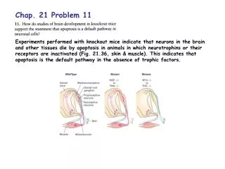
11-3Erythrocyte Abnormalities: Anemia • Anemia - a decrease in oxygen carrying ability, can happen for various reasons such as: • Decrease in RBC number: due to hemorrhage, infection, lack of Vitamin B12, destruction of bone marrow due to cancer, radiation, or certain medications • Inadequate hemoglobin content in RBC’s (iron deficiency anemia) • Abnormal hemoglobin in RBC’s (sickle cell anemia) http://www.youtube.com/watch?v=R4-c3hUhhyc • http://www.pbs.org/wgbh/evolution/library/01/2/l_012_02.html Sickle cell anemia
11-3 Hematopoiesis • Because RBC’s are anucleate, they can’t make proteins, grow, or divide. As they age, they become more rigid and begin to fall apart in about 100 to 120 days, when they become eliminated by phagocytes in the spleen, liver, and other body tissues • Hemoglobin decomposes into iron (Fe2+) and a green pigment called biliverdin • Fe2+ is reused by bone marrow, some biliverdin converted to orangish bilirubin by the liver and is excreted as bile • Hematopoiesis (hemopoiesis) – blood cell formation • Occurs in red bone marrow; each type is produced in different numbers in response to changing body needs and stimuli. After maturation, they are discharged into the blood
11-3 Hematopoiesis (RBC’s) • All formed elements arise from a common type of stem cell, the hemocytoblast, which resides in the red bone marrow. • Their development differs and once a cell is committed to a specific pathway it cannot change. • The hemocytoblastforms two types of descendants: the lymphoid stem cells, which produce lymphocytes and the myeloid stem cell, which can produces all other classes of formed elements.
11-3 Hematopoiesis (RBC’s cont.) • Lost cells are replaced by division of hemocytoblasts in the red bone marrow. • The developing RBCs divide many times and then begin synthesizing huge amounts of hemoglobin. • Suddenly, when enough hemoglobin has been accumulated, the nucleus and most organelles are ejected and the cell collapses inward, resulting in an immature RBC, still containing RER • These enter bloodstream and within 2 days eject remaining RER to become full-functioning RBC’s
11-3 More Hematopoiesis, Hooray! • The rate of erythrocyte production is controlled by a hormone called erythropoietin; produced in kidneys; numbers increase when blood oxygen levels decline; target the red bone marrow • Formation of white blood cells (WBC’s): certain stimulating factors and interleukins prompt red bone marrow to turn out leukocytes and also marshal up an army of WBCs to ward off attacks. • They are released in response to specific chemical signals in the environment. • Platelets: thrombopoietin accelerates production.
11-4 Blood Groups • The plasma membranes of RBCs bears genetically determined surface proteins (A, B, and Rh), which identify each person as unique • Antigen: substance that the body recognizes to release antibodies or use other means to mount an immune defense against it • Binding of antibodies to the antigens causes the RBCs to clump, called agglutination, which leads to clogging of small blood vessels throughout the body and hemolysis.
11-4 Blood Groups (cont.) 46.1% 38.8% 11.1% 3.9%
11-4 Blood Groups (cont.) BLOOD TRANSFUSIONS VIDEO Universal Donor O A B AB Universal Recipient
11-5 The Formed Elements: Leukocytes (WBC’s) • About 1 WBC for every 1000 RBC’s • Make up less than 1% of total blood volume • Large and contain nuclei and other organelles • Have ability to slip in and out of blood vessels, diapedisis. Can also locate areas of tissue damage and infection in the body by responding to certain chemicals that diffuse from the damaged cells, called positivechemotaxis
11-5 Leukocytes (cont.) • WBC’s move through tissue space using ameboid motion • By following the diffusion gradient, they pinpoint areas of damage and rally in large numbers to destroy the microorganisms or dead cells. • When WBC’s move into action, your body increases the numbers rapidly • They are classified into two groups, granulocytes and agranulocytes, depending on whether or not they have visible granules
11-5 Classification of Leukocytes Neutrophils • Granulocytes: have granules, lobed nuclei. • Neutrophils: multi-lobed nucleus; cytoplasm stains pink, avid phagocytes at sites of acute infection • Eosinophils: blue-red nucleus, large brick-red granules, numbers increase during allergies and infections from parasitic worms (tapeworm) – attack antibody labeled substances • Basophils: rare, contain large histamine (promote inflammation) containing granules that stain dark blue Eosinophils Basophils
11-5 Classification of Leukocytes • Agranulocytes: lack visible cytoplasmic granules; normal nuclei • Lymphocytes: have large, dark purple nucleus that occupies most of the cell; play an important role in immune response • Monocytes: largest WBC’s; kidney-bean shaped nucleus; important in fighting chronic infections, such as tuberculosis Lymphocytes Monocytes
11-6 The Formed Elements:Platelets • Fragments of multinucleate cells called megakarocytes, which pinch off platelet pieces, which seal themselves off from the surrounding fluid • Small, darkly staining, irregularly shaped bodies • Normal count is about 300,000/mm3 • Needed for the clotting process that occurs in plasma when blood vessels are ruptured or broken Tiny dots are thrombocytes (t)
11-7 Hemostasis • Hemostasis is the stopping of blood flow; it involves many substances present in plasma, as well as some that are released by platelets and injured tissue cells; involves 3 major phases, which occur in a rapid sequence. • They can be seen in motion, here… http://www.mhhe.com/biosci/esp/2002_general/Esp/folder_structure/tr/m1/s7/trm1s7_3.htm
11-7 Steps of Hemostasis • 1.Vascular spasms occur: the factors causing vessel spasms include direct injury to the smooth muscle cells and stimulation of local pain receptors which causes the blood vessels to go into spasms; the spasms narrow the blood vessel at that point, decreasing blood loss until clotting can occur. • Other factors causing vessel spasms include platelets releasing serotonin
11-7 Steps of Hemostasis • .2.Platelet plug forms: platelets are repelled by an intact endothelium, but when it is broken so that the underlying collagen fibers are exposed, the platelets become sticky and cling to the damaged site. The anchored release chemicals that attract more platelets to the site, and as more pile up, the platelet plug is formed
11-7 Steps of Hemostasis • 3.Coagulation events occur in 3 Pathways EXTRINSIC PATHWAY • The injured tissues release tissue factor (TF), which plays a role in clotting. The more tissue factor released, the faster the clot. • PF3, a phospholipid, coats the platelets, interacts with TF, Vit. K, other clotting factors, and Ca2+, to activate the clotting cascade INTRINSIC PATHWAY • Platelets release platelet factor which combines with Ca2+ and other clotting factors to activate clotting cascade COMMON PATHWAY • This prothrombin activator converts prothrombin, present in the plasma, to thrombin, an enzyme • Thrombin converts fibrinogen to fibrin which forms the clot
11-7 Hemostasis (cont.) • Normally, blood clots within 3-6 minutes; once the cascade begins, the triggering factors are rapidly inactivated, so to prevent widespread clotting. • Clot retraction occurs after clot has been formed, pulls edges of wound closer together making it easier to repair • Applying gauze and pressure to the wound greatly aid the clotting process.
11-7 Blood Disorders • Thrombus: a clot that develops and persists in an unbroken blood vessel • Embolus: a clot that broke away from the vessel wall and floats freely in the bloodstream • Hemophilia: Hereditary; several different disorders resulting from a lack of any substance needed for clotting; get synthetics injected into them to stop the bleeding CHOLESTEROL IN BLOOD VIDEO
Sources • http://uhaweb.hartford.edu/BUGL/RBCs.jpg • http://www.thecrookstoncollection.com/Collection/medslides/Slides/CGL-Buffy-Coat.jpg • http://www.dreddyclinic.com/images/ph_scale.jpg • http://www.uoguelph.ca/zoology/devobio/miller/013633fig6-14.gif • http://www.uoguelph.ca/zoology/devobio/miller/013631fig6-15.gif • http://www.sciencemuseum.org.uk/exhibitions/genes/images/1-3-5-1-4-2-1-3-1-0-0.jpg • http://www.wadsworth.org/chemheme/heme/microscope/pix/cmlmyeloblast_nw.jpg • http://www.ctdslab.co.uk/images/felineneutlarge.jpg • http://www.funsci.com/fun3_en/blood/blood_03.jpg • http://cellbio.utmb.edu/microanatomy/blood/basophil.JPG • http://mmserver.cjp.com/images/image/4303037.jpg • http://mmserver.cjp.com/images/image/0144088.jpg • http://www.wadsworth.org/chemheme/heme/microscope/pix/platelets_nw.jpg • http://content.answers.com/main/content/wp/en/thumb/b/b8/180px-Gray72.png • http://content.answers.com/main/content/wp/en-commons/2/20/Illu_blood_cell_lineage.jpg • http://www.sciencecases.org/baby_joe/hematopoiesis.gif • http://www.hemophiliagalaxy.com/patients/images/REC0057.jpg • http://www.ncbi.nlm.nih.gov/books/bookres.fcgi/stryer/ch10f37.gif • http://www.made-in-china.com/image/2f0j00fIRETCQPjaKMM/Conforming-Elastic-Bandage-Gauze-.jpg