Download
1 / 42
580 likes | 1.53k Views
Coagulation failure in pregnancy. Dr : Hashmi Hajrai MBBCh , DGO, M’MAS, MRCOG Consultant Obstetrician & Gynaecologist. Learning objectives. The student should understand the alterations in coagulations & fibrinolysis associated with pregnancy

E N D
Coagulation failure in pregnancy Dr : HashmiHajrai MBBCh, DGO, M’MAS, MRCOG Consultant Obstetrician & Gynaecologist
Learning objectives • The student should understand the alterations in coagulations & fibrinolysis associated with pregnancy • Refresh his mind about the normal coagulation cascade mechanisms and its triggers • Broad line classification of coagulation failure in pregnancy
Understanding the pathogenesis of DIC syndrome, diagnosis, complications & management outlines • Brief knowledge on some other important causes of coagulation failure in pregnancy
Coagulations changes in pregnancy • Bleeding during labour is dealt with effectively by - increased production of coagulation factors during pregnancy - increased blood volume - myometrial contraction
this hypercoagulable state with local activation of clotting system is associated with increased risk of not only VTE but also DIC
The fibrinolytic system is responsible for disposing of fibrin after fulfilling its haemostatic function • Plasma proteases are responsible for controlling the speed and extent of coagulation & fibrinolysis
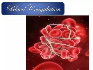
HEMOSTASISPrimary + Secondary + Tertiary • Primary Hemostasis • Platelet Plug Formation:dependent on normal platelet number & function • Secondary Hemostasis • Activation of Clotting Cascade Deposition & Stabilization of Fibrin • Tertiary Hemostasis • Dissolution of Fibrin Clot:dependent on Plasminogen Activation
Normal Artery Endothelium Smooth Muscle Adventitia
Second step is activation of coagulation Three phases • Intrinsic pathway • Extrinsic pathway • Common pathway
Coagulation cascade Intrinsic pathway XII XI Extrinsic pathway IX VII APTT VIII X PT thrombin Prothrombin (II) V, Ca, P/L fibrin fibrinogen XIII STABILISED FIBRIN
Classification of coagulation disorders Congenital coagulation failure disorders these are uncommon.....examples: • Von Willebrand’s disease...will be discussed • Haemophilia A & B
Acquired coagulation failure disorders are far more commonly seen • Thrombocytopenic coagulopathies • Disseminated intravascular coagulation ..DIC • Anticoagulant therapy
4. Congenital Coagulopathies Von Willebrand disease Factor synthesized by endothelial cells & megakaryocytes Forms a complex with factor VIII Mediates platelet adhesion and collagen Inherited as autosomal dominant trait
Congenital Coagulopathies Von Willebrand disease During pregnancy Prophylactic treatment factor VIII level below 25% DDAVP is administered as labor begins – repeated every 12 hrs. FFP or cryoprecipitate (500-1,500 units of factor VIII activity)
Congenital Coagulopathies Von Willebrand disease During labor Factor VIII levels should be maintained at 50% of normal CS – factor VIII level to 80%of normal Check daily during the post partum period
Congenital Coagulopathies Other coagulation factor deficiencies Factor VIII ( hemophilia A) Factor IX ( hemophilia B)
Thrombocytopenic Coagulopathies Autoimmune Thrombocytopenic Purpura Idiopathic thrombocytopenic purpura Immunoglobulin G (IgG)
Thrombocytopenic Coagulopathies Diagnosis Platelet count < 100,000/mm3 Increased numbers of megakaryocytes Increased platelet volume Diameter
Thrombocytopenic Coagulopathiestreatment Conservative management Corticosteriods – if platelet count <20,000/mm3 before the onset of labor or < 50,000/mm3 at time of delivery High dose IV immunoglobulin produces increase in platelet count Significant hemorrhage – immediate postpartum period platelet transfusion
The theoretical risk of intracranial haemorrhage in the thrombocytopenic foetus has not been shown to be reduced by C/S therefore C/S should be performed for obstetric reasons
DIC SYSTEMIC ACTIVATION OF COAGULATION • An acquired syndrome characterized by systemicintravascularcoagulation • Coagulation is always the initial event Intravascular deposition of fibrin Depletion of platelets and coagulation factors Thrombosis of small and midsize vessels Bleeding DEATH Organ failure
Obstetric causes of DIC Falls into three categories • conditions associated with release of tissue thromboplastin that activates extrinsic pathway - placental abruption - dead foetus - molar pregnancy • Conditions associated with endothelial damage leading to activation of intrinsic & extrinsic pathways - pre-eclampsia & eclampsia
Conditions having non-specific or indirect action - amniotic fluid embolism - gram negative septicaemia - saline abortion
Mechanism of DIC Bick et al., 2002
Clinical manifestation of DIC • Those of the underlying cause • Those due to Complications of DIC
Haemorrhagic manifestations Involving skin & mucus membranes • Ecchymosis • Petechiae • Bleeding from the gum • Haematuria • GIT bleeding • Venepunctur oozing • Intracranial or intracerebral haemorrhage
Thrombotic manifestations • Neurologic with multifocal lesions , delirium & coma • Dermatologic with focal ischaemia & superficial gangreen • Renal with cortical necrosis and ureamia • GIT acute ulceration with bleeding • Vascular occlusion causing pulmonary infarction or peripheral vascular gangreen
Lab results • Markedly decreased platelet count • Markedly Increased fibrin degradation products FDP’s • Fragmented RBCs & microspherocytes in peripheral blood film • Low fibrinogen , factor II , V & VII • Prolonged PT, PTT & TT
Microscopic findings in DIC • Fragments • Schistocytes • Paucity of platelets
Fragmented RBC T. TATU
Treatment of DIC • Remove underlying cause • Replenish depleted factors • FFP Provides source of most factors • Cryoprecipitate provides fibrinogen • Platelet and blood support • Cautious use of heparin Up to date, emedicine
conclusion • Blood coagulation is a major component of haemostasis. Increased Coagulation factors levels in pregnancy is meant to minimize blood loss at time of delivery • This haemostatic mechanism could fail risking patient’s life
Thrombocytopenic coagulation failure and DIC syndrome are the most commonly seen in obstetric practice • Congenital causes of coagulation failure are uncommon and usually already diagnosed prior to pregnancy • DIC syndrome is always secondary to an underlying pathology
If diagnosis of DIC is missed or appropriate action is delayed it can cause serious maternal morbidity or even death • Platelet transfusion and coagulation factor replacement or fresh blood transfusion are the main stay of treatment besides other supportive therapy
Use of heparin is controversial . Haematologist opinion should be sought before it’s use