Download
1 / 73
910 likes | 1.71k Views
Septic Shock and Disseminated Intravascular Coagulation in Pregnancy. DIC. Consumptive coagulopathy a pathological activation of coagulation (blood clotting) mechanisms that happens in response to a variety of diseases

E N D
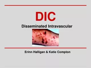
Septic Shock and Disseminated Intravascular Coagulation in Pregnancy
DIC • Consumptive coagulopathy • a pathological activation of coagulation (blood clotting) mechanisms that happens in response to a variety of diseases • a situation of inappropriate coagulation within the blood vessels which leads to the consumption of clotting factors, thus resulting in the failure of the clotting mechanism at the site of bleeding • leads to the formation of small blood clots inside the blood vessels throughout the body
DIC • As the small clots consume coagulation proteins and platelets, normal coagulation is disrupted and abnormal bleeding occurs • The small clots also disrupt normal blood flow to organs such as the kidneys • can occur acutely but also on a slower, chronic basis, depending on the underlying problem • common in the critically ill, and may participate in the development of multiple organ failure, which may lead to death
DIC • Begins with an event that triggers widespread clotting with the formation of microthrombi throughout the circulation • Triggers fibrinolysis, which is the bodies’response to the abnormal clotting by attempting to break up the unneeded clots • Production of FDPs that further reduce the efficiency of normal clotting process
If DIC occurs during or after delivery, the reduced level of clotting factors and the presence of FDPs prevent normal hemostasis at the placental site • FDPs inhibit myometrial action and prevent the uterine muscle from constricting the blood vessels in a normal way • Torrential hemorrhage may be the outcome, and even if clotting does occur, the clot is unstable • Microthrombi in the bloodstream may cause circulatory obstruction in the small blood vessels and lead to cyanosis of fingers and toes to CVAs, or organ failure
Abruptio Placenta • Premature separation of the normally implanted placenta • Occurs in 1 in 200 deliveries • Can cause concealed or external hemorrhage
Abruptio Placenta • Concealed hemorrhage • Effusion of blood behind the placenta but with intact margins • Completely separated placenta with membranes still attached to the uterine wall • Blood gains access to the amniotic cavity after breaking through the membranes • Fetal head closely applied to the lower uterine segment that the blood cannot make its way past it
Abruptio Placenta • Symptoms: • Vaginal bleeding • Hypertonic uterus that is tender on palpation • Fetal heart rate deceleration
Abruptio Placenta • Risk Factors: • Increased age and parity • Preeclampsia/ chronic HPN • PROM • Multifetal gestation • Hydramnios • Cigarette smoking • Prior abruption • External trauma • Leiomyomas
Abruptio Placenta • Pathology • Hemorrhage into the decidua basalis • The decidua splits leaving a thin layer adherent to the myometrium • Development of decidual hematoma that leads to separation, compression, and the ultimate destruction of the placenta adjacent to it • Rupture of decidual spiral artery causing a retroplacental hematoma
Abruptio Placenta • Bleeding is almost always maternal • Significant fetal bleeding occurs in traumatic abruption which results from a tear or fracture in the placenta rather than from the placental separation
Abruptio Placenta • Complications: • Shock – occurs in proportion to blood loss • Uteroplacental apoplexy • Couvelaire uterus • Widespread extravasation of blood into the myometrium and serosa • Not an indication for hysterectomy
Abruptio Placenta • Complications: • DIC • Placental abruption causes damaged tissue at placental site and large quantities of thromboplastins are released into the circulation, resulting in large scale clotting throughout the system, not just placental site • Overt hypofibrinoginemia with increase levels of fibrinogen-fibrin degradation products • Fibrin may in turn cause small blood vessel occlusion resulting in tissue necrosis, occuring more often in the glomerular capillaries causing acute renal failure
Abruptio Placenta • Management • Nasal oxygen • IV hydration • Prepare blood for possible transfusion • Evaluate hematologic and clotting studies (CBC, PT, aPTT, Fibrinogen, platelet count) • Monitor urine output • Continuous fetal heart rate monitoring • Amniotomy • Delivery of the baby
Abruptio Placenta • Management • Delivery of the baby • Vaginal delivery – if the fetus is dead and the mother is hemodynamically stable and with controlled vaginal bleeding • Cesarean delivery Evidence of fetal compromise Severe uterine hypertonus Life threatening vaginal bleeding DIC when vaginal delivery is not imminent
Intrauterine Fetal Death • May be secondary to abortion, abruptio placenta or other pregnancy-related complications • Consumptive coagulopathy usually occurs when the dead fetus is retained in utero for 4weeks or more • Hypofibrinogenimia with increase serum fibrin degradation products with or without decrease platelet count
Intrauterine Fetal Death • retained fetus of more than 3 or 4 weeks causes thromboplastins to be released from the fetal tissue, into the maternal circulation, causing the onset of clotting problems • widespread clotting with the formation of microthrombi throughout the circulation • triggers fibrinolysis and FDP production • The FDPs reduce the efficiency of normal clotting
Intrauterine Fetal Death • Management • Delivery of the dead fetus • Correction of hematologic and clotting problems • Blood transfusion • Antibiotics
Preeclampsia • Pregnancy-specific syndrome of reduced organ perfusion secondary to vasospasm and endothelial activation • Minimum criteria: • BP>/= 140/90 after 20 weeks AOG • Proteinuria >/= 300mg/24hrs or >/= +1 dipstick
Preeclampsia • Increase severity of preeclampsia • DBP >/= 110 mmHg • Proteinuria >/= +2 dipstick • + headache, visual disturbances, upper abdominal pain • Elevated liver enzymes and serum creatinine • Thrombocytopenia < 100,000/mm3 • Pulmonary edema
Preeclampsia • Epigastric or RUQ pain • Hepatocellular necrosis, ischemia, and edema that stretches the Glisson’s capsule • Hepatic infarction and hemorrhage or catastrophic rupture of a subcapsular hematoma • Accompanied by elevated serum liver enzymes • A sign to terminate pregnancy • Thrombocytopenia • Caused by platelet activation and aggregation as well as microangiopathic hemolysis induced by severe vasospasm
Preeclampsia • DIC and preeclampsia • unknown, and unclear precursor to DIC • patients have higher amounts of FDPs in the blood and urine than others • Thrombocytopenia, increase intravascular coagulation and erythrocyte destruction can contribute to the development of DIC • increase fibrinolysis and increase production of FDPs
Amniotic Fluid Embolism • Characterized by the abrupt onset of hypotension, hypoxia, and consumptive coagulopathy • thought to occur when amniotic fluid , fetal cells, hair, or other debris enter the maternal circulation • Overall incidence ranges from 1 in 8,000 to 1 in 80,000 pregnancies
Amniotic Fluid Embolism • 75 % of survivors are expected to have long-term neurologic deficits. • If the fetus is alive at the time of the event, nearly 70 % will survive the delivery but 50% of the survived neonates will incur neurologic damage.
Risk Factors • Advanced maternal age • Multiparity • Meconium • Cervical laceration • Intrauterine fetal death • Uterine tetanic contractions • Precipitate labor • Placenta accreta • Polyhydramnios • Uterine rupture • Maternal history of allergy or atopy • Chorioamnionitis • Macrosomia • Male fetal sex • Oxytocin (controversial)
Amniotic Fluid Embolism • Pathophysiology • Phase 1: Pulmonary and systemic HPN Amniotic fluid and fetal cells enter the maternal circulation biochemical mediators pulmonary artery vasospasm pulmonary hypertension elevated right ventricular pressure hypoxia myocardial and pulmonary capillary damage left heart failure acute respiratory distress syndrome
Amniotic Fluid Embolism • Pathophysiology • Phase 2 biochemical mediators DIC Hemorrhagic phase characterized by massive hemorrhage and uterine atony.
Amniotic Fluid Embolism • Clinical Presentation (1) Respiratory distress (2) Cyanosis (3) Cardiovascular collapse (cardiogenic shock) (4) Hemorrhage (5) Coma.
Clinical Presentation • A sudden drop in O2 saturation can be the initial indication of AFE during c/s. • More than 1/2 of patients die within the first hour. • Of the survivors 50 % will develop DIC which may manifest as persistent bleeding from incision or venipuncture sites. The coagulopathy typically occurs 0.5 to 4 hours after phase 1.
Amniotic Fluid Embolism • Pathogenesis: • Amniotic fluid enters the maternal circulation as a result of a breach in the physiological barrier that normally exists between maternal and fetal compartments • Amniotic fluid abnormally enters the maternal venoussystem via the endocervical veins, the placental site (if placentais separated), or a uterine trauma site
Amniotic Fluid Embolism • Diagnosis: • Detection of squamous cells or other debris of fetal origin in the central pulmonary circulation • Clinical by identifying characteristic signs and symptoms
Amniotic Fluid Embolism • Management: • Restoration of cardiovascular and pulmonary equilibrium - Maintain systolic blood pressure >90 mm Hg. - Urine output > 25 ml/hr - Arterial pO2 > 60 mm Hg. • Re-establishing uterine tone • Correct coagulation abnormalities There are no data that supports that any type of intervention improves maternal prognosis with amniotic fluid embolism
Amniotic Fluid Embolism • DIC and AFE • Release of thromboplastins from the amniotic fluid into the maternal circulation • Increase fibrinoloysis • Increase FDPs that further impairs the clotting mechanism
Sepsis Syndrome • Induced by a systemic inflammatory response to bacteria or their by-products such as endotoxins or exotoxins • Most commonly due to: • Acute pyelonephritis • Chorioamnionitis • Puerperal infection • Necrotizing fasciitis
Acute Pyelonephritis • shaking chills • fever (T>38oC) • flank pain • nausea and vomiting • with or without signs and symptoms of lower urinary tract infections • Costovertebral angle tenderness
Acute Pyelonephritis • Diagnosis: • Urinalysis: pyuria >/= 5 wbc/hpf of centrifuged urine • Urine culture and sensitivity • Bacteriuria >/= 10,000 cfu of a uropathogen/ml of urine • Escherichia coli – most common isolate (75-80%) • Klebsiella pneumoniae (10%) • Enterobacter or Proteus (10%)
Acute Pyelonephritis • Management: • Hospitalization • Urine culture and sensitivity • CBC, serum creatinine • Empiric treatment with IV antibiotic • Post treatment urine culture
Chorioamnionitis • Denotes histologic infection wherein microorganisms and PMNs reside in the layers between the chorion and the amnion • Applies only to pregnancies in which the fetus has achieved viability, to differentiate it from septic abortion
Chorioamnionitis • Diagnosis: Clinical History – Risk Factors • First pregnancy • Young age • Preterm labor (10x increase risk) • Prelabor rupture of membranes (10x increase risk) • Ruptured membranes >12 hours • Prolonged active phase of labor • Primigravid >12 hours • Multigravid >8 hours • Use of intrauterine monitors
Chorioamnionitis • Diagnosis: Clinical History – Risk Factors • Frequent and numerous vaginal examinations (>6 times) • Presence of meconium in the AF (4x increase risk) • History of untreated or inadequately treated abnormal vaginal flora
Chorioamnionitis • Diagnosis: Physical Examination • Presence of 2 out of 3 clinical signs • Maternal fever – oral temp >37.8oC - hallmark for the diagnosis • Uterine tenderness • Persistent fetal tachycardia
Chorioamnionitis • Treatment • Antimicrobial therapy • Should be given at the time of diagnosis • Ampicillin is the drug of choice for fetal therapy • Aminoglycoside is given as maternal therapy to prevent development of septic shock from Enterobacteriaceae • If foul smelling amniotic fluid is present, give Metronidazole for anaerobic coverage
Chorioamnionitis • Treatment • Delivery • Chorioamnionitis is an indication for delivery but is not an indication for cesarean birth • CS is indicated if there is fetomaternal complications or in the presence of obstetric indications