Download
1 / 48
550 likes | 693 Views
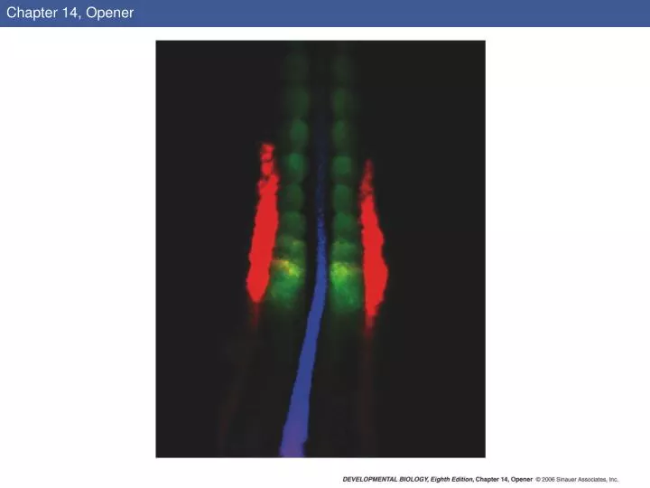
Chapter 14, Opener. 14.1 The major lineages of the amniote mesoderm (Part 1). 14.1 The major lineages of the amniote mesoderm (Part 2). 14.2 Gastrulation and neurulation in the chick embryo, focusing on the mesodermal component (1).

E N D
14.2 Gastrulation and neurulation in the chick embryo, focusing on the mesodermal component (1)
14.2 Gastrulation and neurulation in the chick embryo, focusing on the mesodermal component (2)
14.4 Neural tube and somites seen by scanning electron microscopy
14.6 Somite formation correlates with the wavelike expression of the hairy1 gene in the chick
14.7 Possible scheme for the regulation of the clock through which an Fgf8 gradient regulates a Wnt oscillating clock, which in turn controls a Notch clock
14.8 Ephrin and its receptor constitute a possible cut site for somite formation (Part 1)
14.8 Ephrin and its receptor constitute a possible cut site for somite formation (Part 2)
14.9 Epithelialization and de-epithelialization in somites of a chick embryo
14.10 Segmental plate mesoderm transplanted into a different region in a younger embryo differentiates according to its original position
14.11 Diagram of a transverse section through the trunk of a chick embryo on days 2–4 (Part 1)
14.11 Diagram of a transverse section through the trunk of a chick embryo on days 2–4 (Part 2)
14.12 Model of major postulated interactions in the patterning of the somite
14.13 Conversion of myoblasts into muscles in culture (Part 1)
14.13 Conversion of myoblasts into muscles in culture (Part 2)
14.13 Conversion of myoblasts into muscles in culture (Part 3)
14.14 Schematic diagram of endochondral ossification (Part 1)
14.14 Schematic diagram of endochondral ossification (Part 2)
14.16 Skeletal mineralization in 19-day chick embryos that developed (A) in shell-less culture and (B) inside an egg during normal incubation
14.17 Respecification of the sclerotome to form each vertebra
14.18 Scleraxis is expressed in the progenitors of the tendons
14.19 Induction of scleraxis in the chick sclerotome by Fgf8 from the myotome (Part 1)
14.19 Induction of scleraxis in the chick sclerotome by Fgf8 from the myotome (Part 2)
14.19 Induction of scleraxis in the chick sclerotome by Fgf8 from the myotome (Part 3)
14.20 Signals from the paraxial mesoderm induce pronephros formation in the intermediate mesoderm of the chick embryo (Part 1)
14.20 Signals from the paraxial mesoderm induce pronephros formation in the intermediate mesoderm of the chick embryo (Part 2)
14.21 General scheme of development in the vertebrate kidney (Part 1)
14.21 General scheme of development in the vertebrate kidney (Part 2)
14.22 Reciprocal induction in the development of the mammalian kidney
14.24 Ureteric bud growth is dependent on GDNF and its receptors (Part 1)
14.24 Ureteric bud growth is dependent on GDNF and its receptors (Part 2)
14.26 Lim1 expression (dark stain) in a 19-day embryonic mouse kidney
14.27 The effect of GDNF on the branching of the ureteric epithelium
14.28 Signaling molecules and branching of the ureteric epithelium
14.29 Development of the bladder and its connection to the kidney via the ureter (Part 1)
14.29 Development of the bladder and its connection to the kidney via the ureter (Part 2)
14.29 Development of the bladder and its connection to the kidney via the ureter (Part 3)