1 / 36

Chapter Opener 17

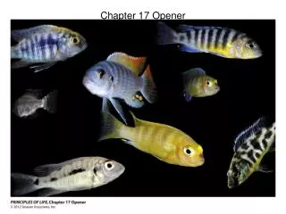
Chapter Opener 17. Figure 17.1 The major components of whole blood. Table 17.1 Composition of Plasma (1 of 2). Table 17.1 Composition of Plasma (2 of 2). Figure 17.2 Photomicrograph of a human blood smear stained with Wright’s stain.

trung
Download Presentation

Chapter Opener 17

An Image/Link below is provided (as is) to download presentation Download Policy: Content on the Website is provided to you AS IS for your information and personal use and may not be sold / licensed / shared on other websites without getting consent from its author. Content is provided to you AS IS for your information and personal use only. Download presentation by click this link. While downloading, if for some reason you are not able to download a presentation, the publisher may have deleted the file from their server. During download, if you can't get a presentation, the file might be deleted by the publisher.

E N D

Presentation Transcript


  1. Chapter Opener 17

  2. Figure 17.1 The major components of whole blood.

  3. Table 17.1 Composition of Plasma (1 of 2)

  4. Table 17.1 Composition of Plasma (2 of 2)

  5. Figure 17.2 Photomicrograph of a human blood smear stained with Wright’s stain.

  6. Figure 17.3 Structure of erythrocytes (red blood cells).

  7. Figure 17.4 Structure of hemoglobin.

  8. Figure 17.4a Structure of hemoglobin.

  9. Figure 17.4b Structure of hemoglobin.

  10. Figure 17.5 Erythropoiesis: formation of red blood cells.

  11. Figure 17.6 Erythropoietin mechanism for regulating erythropoiesis.

  12. Figure 17.7 Life cycle of red blood cells.

  13. Figure 17.7 Life cycle of red blood cells. (1 of 2)

  14. Figure 17.7 Life cycle of red blood cells. (2 of 2)

  15. Figure 17.8 Sickle-cell anemia.

  16. Figure 17.8a Sickle-cell anemia.

  17. Figure 17.8b Sickle-cell anemia.

  18. Figure 17.9 Types and relative percentages of leukocytes in normal blood.

  19. Figure 17.10 Leukocytes.

  20. Figure 17.10a Leukocytes.

  21. Figure 17.10b Leukocytes.

  22. Figure 17.10c Leukocytes.

  23. Figure 17.10d Leukocytes.

  24. Figure 17.10e Leukocytes.

  25. Figure 17.11 Leukocyte formation.

  26. Table 17.2 Summary of Formed Elements of the Blood (1 of 2)

  27. Table 17.2 Summary of Formed Elements of the Blood (2 of 2)

  28. Figure 17.12 Formation of platelets.

  29. Figure 17.13 Events of hemostasis.

  30. Table 17.3 Blood Clotting Factors (Procoagulants)

  31. Figure 17.14 The intrinsic and extrinsic pathways of blood clotting (coagulation).

  32. Figure 17.14 The intrinsic and extrinsic pathways of blood clotting (coagulation). (1 of 2)

  33. Figure 17.14 The intrinsic and extrinsic pathways of blood clotting (coagulation). (2 of 2)

  34. Figure 17.15 Scanning electron micrograph of erythrocytes trapped in a fibrin mesh.

  35. Table 17.4 ABO Blood Groups

  36. Figure 17.16 Blood typing of ABO blood types.

More Related