Download
1 / 39
850 likes | 2.82k Views
Protein Trafficking. Vesicle transport and targeting in the secretory pathway COP coated vesicles SNAREs Protein sorting Secretion - Golgi to plasma membrane Retention in ER Golgi to lysosome. Protein Trafficking - Regulated transport to the trans-Golgi network.

E N D
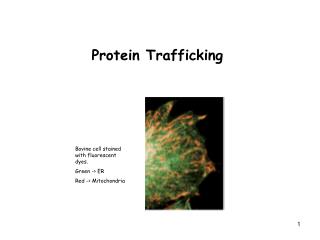
Protein Trafficking Vesicle transport and targeting in the secretory pathway COP coated vesicles SNAREs Protein sorting Secretion - Golgi to plasma membrane Retention in ER Golgi to lysosome
Protein Trafficking - Regulated transport to the trans-Golgi network • Multimeric proteins (e.g., ion channels). - KATP channels = 4 Kir6.1/6.2 subunits with 4 SUR1/2A/2B subunits in ER. - NMDAR = combination of NR1, NR2A-D, or NR3A-B subunits. - GABAAR: 16 different mammalian isoforms (α1-6, β1-3, γ1-3, δ, ε, π, and θ), making the total number of receptor combinations = 165; but only ~20-30 functionally distinct receptor types exist.
During GABAAR assembly, chaperones, IgG-bp (BiP) and calnexin, interact with subunits. • Association with ER depends on ER retention signals (KDEL). • Hydrophobic residues. • Exact mechanism of ER retention involves interaction with ER matrix, failure to be recruited for transport, or retrieved from the cis Golgi. • Coatomer proteins (COPS) are involved in the selection of cargo for anterograde (COPII) or retrograde (COPI) transport between organelles.
Morphology • Diffusion barrier • Cytoskeleton = actin-spectrin-ankyrin anchors membrane proteins (e.g., Nav channels). • High [protein] crowding. • Soma, dendrites, axon - not 1 continuous structure. • Inhibitory synapses vs excitatory synapses.
Development of Polarity • Synapse Formation: - GFP-PSD-95 visualized extension and maintenance of filopodia. - Appeared to be translocated to filopodia as pre-assembled clusters, rather than as accumulating gradually. - But occurs only when the postsynaptic scaffold/signaling complex is already there. - Within 45 min, AMPA and NMDA receptors can be found postsynaptically.
Development of Polarity • Axonal Development - Nav channels cluster at the Nodes of Ranvier. - Mechanism of how this occurs is unknown. - In demyelinated axons, some form of anchoring occurs via Ankyrin G within the axon. -In the PNS, paranodalKv channels appear to cluster initially within nodes prior to lateral diffusion to their final destination.
Development of Polarity Dendrite + - - + Axon - - + + mGluR2 mGluR7
Polarity Signals • Dendrites hydrophobic motifs • Axons – GAP43
Postysynaptic Targeting • mRNA Targeting • Protein targeting via lipid rafts • Specific transport pathways and proteins - GABAA receptors - NMDA receptors - AMPA receptors
Transport between organelles is mediated by coated vesicles Clathrin coated vesicles mainly involved in endocytosis COP coated vesicles mediate ER to Golgi and back
Transport between ER and Golgi compartments occurs via “COP-coated vesicles”… • Collection of 4-7 “coat proteins” = “COPs”…(aka “Coatomers” ) • COP-coated vesicles function in transport between: • ER and Golgi • Golgi and ER (retrieval) • intra-Golgi • TGN and plasma membrane
COP proteins Cop coated vesicles contain many proteins More COP proteins “cargo” Lipid bilayer Sar1 COPII-coated vesicles - ER to Golgi- SarI in ER membrane COPI coated vesicles - Golgi to ER ARF (instead of Sar1) in Golgi membrane We will only consider Sar1
Sar1 ARF triggers vesicle formation Sar1: GTPase switchon/off ON: binds membranerecruits COP proteins COP proteins then recruit specific cargo Sar1 -- Similar to RAN in nuclear import
Pi GDP Sar1 GTPase GTP GTP Sar1 GTPase “on” “off” GDP GTPase (GTP Binding Proteins) Large family (Ras) of proteins Molecular “switches” GAP In cytoplasm, large amount in “off” form Bound tomembrane GEF cytoplasmic
Sar1 activation exposes hydrophobic tail and membrane insertion Greasy foot Sar 1 in membrane recruits COP proteins
The Ras “superfamily” of small GTPases… • Ras: signaling and regulating cell proliferation… >30% of human tumors have Ras mutations… Many (not all) Ras family members associated with membranes via covalent fatty acid tail (“greasy feet”)… • EF-1/EF-Tu: translation… • Ran: nuclear transport… • Rho family (Rho, Rac, cdc42): actin assembly and organization • Arf/Sar family of “Coat recruitment GTPases:” COP assembly and vesicle budding… • Rab family: vesicle targeting and fusion (see below)
GTP GDP + Pi A A + B B Bound Unbound Cells make high-affinity transient molecular complexes as trigger or switch Aside: G-proteins and ATPases as molecular switches • A paradox: • High-affinity/high-specificity = stable… • Energy input is required to dissociate high-affinity complexes… • (Example: to remove Sar 1 from membrane) • Polymer dynamics: • Actin (ATP), Tubulin (GTP) • Dynamin (GTP) • Motors: • Myosin (ATP), Dynein (ATP) • Kinesin (ATP) • Signaling: • Heterotrimeric G proteins (GTP) • Ras family (GTP) • Translation: • IFs (GTP), EF-1/EF-Tu (GTP) • EF-2/EF-G (GTP) • Chaperones: • HSP70 family (ATP) • HSP60 (ATP) • SRP family: • SRP54 (GTP), SRP-Ra (GTP) • SRP-Rb (GTP)
COP subunits recruit specific cargo proteins… Summary of COPII-coated vesicle formation
Target compartment 1. Formation of coated buds… Coat proteins (“COPs”) Donor compartment Vesicle transport is a complex process 2. Formation of coated transport vesicle… 3. Targeting and docking to specific compartment… SNAREs and Rabs (ATP, GTP, and cytoplasmic protein factors…)
Budding Uncoating, targeting and docking t-SNAREs v-SNAREs Cargo The Snare hypothesis: v- and t-SNAREs target transport vesicles to the correct membrane Specific pairing of V-SNAREs with T-SNAREs matches vesicle to target membrane compartment (>20 known snares in animals cells) Targeting and docking requires/is facilitated by specific RabGTPase in vesicle and Rabeffector in target (~30 known Rabs in animal cells)…
Bacterial toxins target the vesicle docking and fusion machinery of neurons A small subunit of the toxin acts as a specific protease that cleaves and inactivates targeting proteins SNAP25 (t-SNARE) VAMP (v-SNARE) Syntaxin (t-SNARE) VAMP (v-SNARE) Botulism A Botulism B Botulism C Tetanus Net result is to block neuronal signaling by blocking neurotransmitter release (regulated secretory pathway)
Target compartment GTP 4. Uncoating… GDP + Pi Donor compartment GTPgS Vesicle transport is a multi-step process 2. Formation of coated transport vesicle… 3. Targeting and docking to specific compartment… SNAREs and Rabs (ATP, GTP, and cytoplasmic protein factors…) 1. Formation of coated buds… Sar 1 Coat proteins (“COPs”) GTPgS and other non-hydrolyzable GTP analogs block uncoating, resulting in accumulation of docked, coated vesicles GTP hydrolysis by Sar1 is required for uncoating
Target compartment GTP 4. Uncoating… GDP + Pi Donor compartment Vesicle transport is a multi-step process 2. Formation of coated transport vesicle… 3. Targeting and docking to specific compartment… SNAREs and Rabs (ATP, GTP, and cytoplasmic protein factors…) 1. Formation of coated buds… Sar1 GEF and Sar1 Coat proteins (“COPs”) GEF in donor membrane promotes nucleotide exchange, activating Sar1 @ ER, (ARF @ Golgi) and promoting coat assembly… GTP hydrolysis serves as “timer” delaying uncoating (GAP in target membrane?)… GTPase “cycle” provides directionality to vesicle coating/uncoating
Target compartment GTP 4. Uncoating… GDP + Pi Donor compartment 5. Fusion… Vesicle transport is a multi-step process 2. Formation of coated transport vesicle… 3. Targeting and docking to specific compartment… SNAREs and Rabs (ATP, GTP, and cytoplasmic protein factors…) 1. Formation of coated buds… Coat recruitment GTPase GNRP/GEF and Coat recruitment GTPase Coat proteins (“COPs” or “coatomer”) SNARE plus other fusion proteins
SNAREs are necessary for membrane fusion Much still to learn!!! ECB 15-21 SNAREs bring two membranes into close apposition Lipids flow between membranes - fusion Other proteins cooperate with SNAREs to facilitate fusion and to pry SNAREs apart
Vesicle transport and targeting in the secretory pathway COP coated vesicles SNAREs Protein sorting/targeting Secretion - Golgi to plasma membrane Retention in ER Golgi to lysosome How are proteins sorted to appropriate vesicles so that they are transported to proper location? What are the address label?
Two secretory pathways; constitutive and regulated Default pathway for ER/Golgi proteins If no address label, then secrete However, recent data suggests there may be ER exit sequences.. For now, consider secretion default Signal required to trigger secretory granule fusion Example - neurotransmitter release Inside lumen is equivalent to outside of cell secretory_pathway.mov
Regulated secretion Secretory granules containing insulin in pancreatic cells Signal for release is elevated glucose levels in blood
BiP KDEL KDEL-R KKXX They aren’t! Ex: BiP is a member of the HSP70 family that functions in the ER… If secretion is default, how are resident ER proteins retained? Constituitive secretion Secretory granule Regulated secretion Plasma membrane ER CGN C, M, T Golgi TGN Outside BiP escapes from ER and must be “retrieved” from the Golgi… C-terminal KDEL in BiP sequence functions as retrieval signal… KDEL-receptors in Golgi direct retrieval/recycling… KKXX at C-terminus of KDEL-R binds COPI coat and targets back to ER…
Protein targeting RER Golgi ? Vesicletargeting Secretory vesicles Lysosomes Plasma membrane Transport Retrieval Summary so far of protein targeting, revisited… Secretion/membrane proteins Cytoplasm Signal sequence (hydrophobic a-helix) KDEL (soluble proteins) KKXX (membrane proteins) Default Default (regulated secretion) (constituitive secretion) See ECB figure 14-5 How are proteins targeted to the lysosome?
Vesicle transport and targeting in the secretory pathway COP coated vesicles SNAREs Protein sorting Secretion - Golgi to plasma membrane Retention in ER Golgi to lysosome How are proteins sorted to vesicles leaving TGN for lysosome?
Lysosomes degrade and recycle macromolecules… Lysosomes in plant and animal cells contain acid hydrolases (hydrolytic enzymes) for degrading/recycling macromolecules pH of lumen is about 5 - acidic! How are hydrolases and other proteins targeted to lysosomes?
I-cell disease helped decipher the signal for targeting proteins to the lysosome • Recessive mutation in single gene… • Fibroblasts of patients contain large inclusions (I-cells)… • Lysosomes lack normal complement of acid hydrolases… • Alllysosomal enzymes secreted (secretion is the “default” fate for proteins in the ER-Golgi pathway)… • Lysosomal enzymes of “wild-type” (normal) cells are modified by phosphorylation of mannose on oligosaccharide (forming mannose-6-phosphate)… • Lysosomal proteins of I-cells lack M-6-P… • Lysosomal targeting signal resides in carbohydrate!
Clathrin coat RER M6P receptor Cis Golgi Network (CGN) Trans Golgi Network (TGN) Mannose-6-P targets proteins from Golgi to lysosome Transport via clathrin-coated vesicles to… Lysosome Addition of M6P Uncoupling (pH 5) Mature hydrolase Lysosomal hydrolase (precursor) Removal of phosphate & proteolytic processing… M6P receptor recycling back to Golgi Addition of M6P to lysosomal enzymes in cis-Golgi M6P receptor in TGN directs transport of enzymes to lysosome via clathrin-coated vesicles Patients with I-cell disease lack phosphotransferase needed for addition of M-6-P to lysosomal proteins in fibroblasts… secreted…
Postsynaptic Removal of Receptors • Specific endocytotic signals leads to recruitment of AP2 in the internalization of the plasma membrane. • APs recruit clathrin, which instigates membrane invagination and endocytosis. • Examples: - tyr-based signals recruit μ subunits of AP2. - dileu-based signals recruit β subunits of AP2. - Arrestin binding to GPCRs facilitate receptor internalization by its ability to assocociate with clathrin and AP2. - Ubiquitin may recruit AP2 or clathrin, release the receptor from anchoring in the membrane, or recruit receptors to the sites for endocytosis.
AP2 (rapid) EE Trans face Golgi RER LE COPII Lysosome COPI EE AP1 (rapid)
Receptor Endocytosis • Agonist-dependent down-regulation of receptors has been observed for a wide variety of ligands: e.g., GABAA receptors treated with GABA, BDZs, barbs, and neurosteroids; antidepressants and β-adrenergic receptors. • Cell surface receptor number is a balance between 2 competing processes: delivery and removal of receptors. • Synaptic strength is in part, determined by the number of surface AMPA receptors (LTP vs. LTD). BUT… Evidence has shown that in response to NSF-GluR2 interaction, synaptic AMPA receptors are only internalized on the cytoplasmic face of the membrane and are not transported to the soma and degraded in the lysosomes. • Insulin can also cause AMPA receptor down-regulation.
Protein targeting RER Golgi Vesicle targeting Secretory vesicles M6P Plasma membrane Transport Retrieval Protein targeting, revisited Secretion/membrane proteins Cytoplasm Signal sequence (hydrophobic a-helix) Default or signal? KDEL (soluble proteins) KKXX (membrane proteins) Default or signal? (regulated secretion) (constituitive secretion) Lysosomes
The modulation of synaptic strength by alterations in postsynaptic AMPA receptors. Early in development, most of the glu synapses are ‘silent’ at Vm. This results from the presence of NMDA, but not AMPA, receptors in the postsynaptic membrane. Synapses become activated by a NMDA-dep- dent process, leading to the recruitment of AMPA receptors. Synaptic may be incr further, in response to high-freq activity (LTP), by the further recruitment of AMPA receptors.