Download
1 / 44
1.7k likes | 4.58k Views
Hydatid Disease . Dr. Firas Obeidat , MD. Species. Echinococcus granulosis. E. Multilocularis (more malignant). E.vogeli . E.oligarthus . Life - cycle. The major "intermediate host" is a herbivore, mainly the sheep (in other cases is the pig, horse, camel etc).

E N D
Hydatid Disease Dr. FirasObeidat, MD
Species Echinococcus granulosis. E. Multilocularis (more malignant). E.vogeli. E.oligarthus.
Life - cycle The major "intermediate host" is a herbivore, mainly the sheep (in other cases is the pig, horse, camel etc). The major "definitive host", a carnivore mainly the dog ( in other cases is fox, wolves etc). Infection of the intermediate host occurs after ingestion of food contaminated with eggs containing embryos (oncospheres) passed from the faeces of the "definitive host" .
Life - cycle • Humans are infected in two ways: • Direct contact. • Eating products contaminated by the feces of the definitive host.
Morphology • Head (scolex): • 2 rows of hooks • 4 suckers • Body: • 2-6 proglottid
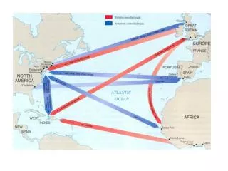
Distribution Endemic areas are Mediterranean countries, Middle East, Turkey, Africa, South America, Asia, Australia and New Zealand. Uncommon in the USA and most of central Europe.
Pathology The eggs (oncospheres) penetrate the wall of the intestine of the intermediate host and via the blood stream they first reach the liver. The liver is affected in (60%), and the lungs in (30%). The right lobe is affected in (80%) and in 1/3 of the cases the cysts are multiple. Single organ involvement in 90% of cases.
Cont. The outer adventitial layer coming from the host is called ectocystorpericyst. The two inner layers coming from parasite, the most inner germinal layer and the outer laminated layer together form the endocyst. The germinal layer secretes the laminated layer which is mucopolysaccharide - protein-lipid complex. The cavity of the cyst contains the hydatid fluid which is clear and similar to interstitial fluid.
Cont. The time required to become mature varies from 10 to 20 months. Daughter cysts (degenerated or secondary cyst), which has fragments of germinal layer. It may develop within the primary cyst or separately. Calcification of the cyst occurs usuylly after 5-10 yrs, commonly in liver cyst . Complete calcification indicate nonviable cyst. Usually protoscolices produced after one year following the infection.
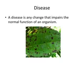
Clinical Presentation Most of the cysts remain uncomplicated (82%). The most common symptom in these cases is right upper quadrant pain, liver enlargement or palpable mass ( 1-5 cm/yr increase in size). Symptoms occur as a result of mechanical effect or generalized toxic reaction due to the presence of parasite itself. Symptoms usually when size is > 10 cm. Suppuration ( 2 bacterial infection) Pressure symptoms.
Clinical Presentation • Rupture into: • The biliary tree (biliary pain, cholangitis, obstjaundice,pancreatitis). • The thorax (bronchpleural fistula). • The peritoneum (anaphylaxis). • The vascular structure ( portal hypertension, Budd chiari syndrome). • GI tract, rare 0.5%.
Clinical Presentation E multilocularis: Liver is the primary site of infection then spreads by direct extension or hematogenous dissemination. It behave like malignancy by invading the tissue affected. 15% multiorgan involvement. If untreated 90% will dye in 10 yrs of developing symptoms.
Diagnosis • Blood test: • In most cases limited eosinophilia < 15% or absent. • In biliary communication: elevated liver enzymes. • Serology for detection of anti-echinococcus antibodies: • Antibody detection is more sensitive than serum antigens (IHA, ELISA). • 85-95% positive for liver cyst and 65% for lung cyst. • IgG ELIZA sensitivity is 94% and specificity is 99%. • More informative in E.multilocularis than E. G. • PCR technology
Diagnosis • Plain X-ray: • Ultrasound: • Simple vs parasitic cyst • Characterestics of hydatid cyst: 1. a collapsing elliptical cyst. 2. detachment of the germinal layer from the cyst wall (water lilly sign). 3. calcification of the rim (eggshell appearance).
Classification upon US findings • Gharbi classification: • type I: a cyst with a pure fluid collection. • type II: fluid collection with split wall (floating • membrane). • type III : fluid collection with septa/daughter cysts • (honeycomb image). • type IV: high internal heterogenous echoes. • type V : a cyst with a calcified, reflecting thick wall.
WHO classification for the functional state of the parasite and size: • Active fertile cysts containing viable scolices. • Transitional compromised by host defence mechanism or by chemotherapy. • Inactive, degenerative, which lost it is fertility. • Small < 5 cm • Medium 5-10 cm • Large > 10 cm
Diagnosis • CT scan: • Sensitivity reaching 100% • Number, size, location, and presence of extrahepatic lesions. • More sensitive in detection of gas and minimal calcifications. • Detect recurrence. • Ultrasound is more informative regarding wall cyst changes. • MRI. • MRCP, ERCP.
Treatment options • Chemotherapy. • Percutaneous drainage. • Surgery: • radical. • conservative. • Approach: • open. • laparoscopic.
Cont. • Chemotherapy: • Albendazole (ABZ) and the active metabolite (after liver metabolism) ABZ sulfoxide is the most effective adjuvant chemotherapy. • better GI absorption. • better tissue distribution. • reaches higher intracystic fluid concentration. • dose: 10-15 mg/kg/day in 2 divided doses. • perioperative regiment most authors agree to a 4 week postoperative period while the preoperative scheme varies from none to 3 months.
Cont. • ABZ alone can lead to a cure rate of 10-30%. • failure rate: in 20-30%. • the WHO recommends the treatment to start somewhere between 1 month and 4 days before surgery.
Cont. A systematic review of the literature published in 2004 concluded that chemotherapy alone is not a successful approach and that it needs to be combined with either percutaneous drainage or surgery which remain the cornerstone of treatment. Dziri C, Haouet K, Fingerhut A. Treatment of hydatid cyst of the liver: where is the evidence? World J Surg 2004; 28 (8):731-6.
WHO recommendations state that medical therapy should be used for: • inoperable disease or unfit patient. • Patients with multiple cysts in two or more organs. • Multiple small liver cysts or cysts deep in liver. • Peritoneal cysts. • Patients following incomplete surgery or relapse. • Prevention of secondary spread of echinococcal infection following spontaneous rupture or aspiration of cysts.
Cont. • Percutaneous drainage: • Some meta-analysis shows that the percutaneous drainage in combination with Albendazole is: • safe and efficient treatment option. • lower complications. • better postoperative recovery. • PAIR and PAIRD (puncture, aspiration, injection, reaspiration, drainage) for large cyst.
Cont. • Indications for PAIR: • > 5 cm in DM. • Infected cyst. • If surgery is contraindicated. • Recurrence after surgery. • Failure of chemotherapy alone. • Pregnant women. • Accessible cysts.
Cont. • Contraindications for PAIR: • Inaccessible or risky location of the cyst. • Extrahepatic location. • Inactive or calcified cyst. • Presence of biliary communications. • undrainable solid material. • Superficial cyst.
Cont. • principles of surgery: • Inactivate the scoleces • avoid spillage. • Eleminate all viable elements of the cyst. • Manage or obliterate the residual cavity. • Indications for surgery: • superficial cyst with risk of rupture. • large cyst > 10 cm with many daughter cyst. • cystobiliary communication. • mass effect to vital organs. • infected cysts. • any extrahepatic localized cyst.
Cont. • Contrindications: • multiple cysts. • Intrahepatic cysts with dificult access. • Dead or totally calcified cysts.
Cont. • Surgical options: • Radical approach: • Pericystectomy. • Cystectomy. • Liver resection. • Less recurrence.
Cont. • Conservative approach: • External drainage. • Wide roof excision. • Evacuation and sterilization of the cavity. • Partail cystopericystectomy. • Near total pericystectomy. • Easier to perform. • Less operative risk. • Higher recurrence rate (10%-30%).
Management of the cavity after conservative approach: • External drainage. • Omentoplasty. • Capitonnage. • Muscle flap (myoplasty).
Cont. • Cosiderations during conservative approach: • Cystobiliary communications: • Presentation. • Preoperative diagnosis and management. • Effect of scolicidal agents. • Management of wide communications: • Biliary bypass. • Sphencteroplasty. • ERCP and ES. • CBD exploration and T tube insertion.
Cont. • Laparoscopic approach vs open: • Contraindications for lap. approach: • Cholangitis due to communication. • Liver cirrhosis. • Complicated hydatid cyst with rupture or infection. • Recurrent cysts. • Deep intraparenchymal cyst. • Cysts located in the posterior segments. • Cysts close to major vessels. • More than 3 cysts. • Thick calcified wall cysts.
Scolicidal agents Peroxide soliution 10% (foaming, gas embolism). Hypertonic saline 10%-20% for 5-10 min. Chlorhexidine 0.05%. Cetrimide 0.4%-1% for 4-10 min. Avoid using these materials in suspected communications, so not to be used routinely.
Postoperative complications: • infection of the residual cavity • intraabdominal abscesses • anaphylactic reactions • spillage of parasite material leading to secondary echinococcosis • biliaryfistulation • sclerosingcholangitis.
CT scan demonstrates apartially calcified multivesicular cyst in the right lobe of the liver.