Download

1 / 72
730 likes | 1.22k Views
Macula defined by anatomists as the macula lutea, or yellow spot containing xanthophyll (yellow) pigment. area with 2 or more layers of ganglion cells that is 5-6 mm in diameter centered vertically between the temporal vascular arcades. Anatomy of macula. Anatomy of macula.

E N D
Macula defined by anatomists as the macula lutea, or yellow spot containing xanthophyll (yellow) pigment. area with 2 or more layers of ganglion cells that is 5-6 mm in diameter centered vertically between the temporal vascular arcades
Anatomy of macula The central 1.5 mm within the macula is occupied by the fovea (or fovea centralis), Within the fovea is a region devoid of retinal vessels known as the foveal avascular zone (FAZ). The geometric center of the FAZ is a central pit known as the foveola, (0.35 mm) Surrounding the fovea is a ring 0.5 mm in diameter, called the parafoveal area where the ganglion cell layer, inner nuclear layer and outer plexiform layer are thickest . Surrounding this zone, a ring approximately 1.5mm wide is termed the perifoveal zone
Macular Edema • Macular edema occurs when fluid and protein deposits collect on or under the macula of the eye a yellow central area of the retina causing it to thicken and swell. The swelling may distort a person's central vision, Cystoid macular edemais a type of macular edema that includes cyst formation.
Macular Edema • Clinically significant macular edema (CSME), is used in diabetic retinopathy • Cystoid macular edema (CME) • Irvine gass syndrum (CME) is used afterafter cataract surgery • Macular cyst: degenerative, Tractional, radiational • Central serous retinopathy (CSR) • Choroidal neovascular membrane (CNVM) • Retinal pigment epithelium detachement (PED)
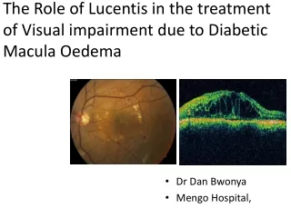
Normal macula cross section as seen with ocular coherence tomography Below – histological cross-section of the macula and underlying choroid
Clinically significant macular edema CSME: Hard exudates at or within 500 u of the center of the macula if associated with thickening of the adjacent retina. CSME: Retinal edema located at or within 500 u of the center of the macula. CSME: A zone of thickening larger than 1 disc area if located within 1 disc diameter of the center of the macula.
Clinically Significant Macular Edema (CSME) is usually associated with diabetes mellitus (DM). It is the most common cause of visual acuity loss with DM
Left – macular thickening from CSME (note the red central zone of the thickness map). Right – normal macular thickness.
Diabetic Macular EdemaMay present as noncystoid or cystoid macular edema
diabetic and hypertensive retinopathy. vitreous traction on the papilla (VPT), and on nasal macula, causing macular oedemaand detachment.
Laser treatments for diabetic retinal changes. White spots represent laser burns. Left – focal treatment. Middle – grid treatment. Right – pan-retinal treatment
Macular Edema • Clinically significant macular edema (CSME), is used in diabetic retinopathy • Cystoid macular edema (CME) • Irvine gass syndrum (CME) is used afterafter cataract surgery • Macular cyst: degenerative, Tractional, radiational • Central serous retinopathy (CSR) • Choroidal neovascular membrane (CNVM) • Retinal pigment epithelium detachement (PED)
Cystoid Macular Edema: Cystoid macular edema (CME) is characterized by intraretinal edema contained in honeycomb-like cystoid spaces. Fluorescein angiography shows the source of edema to be abnormal perifoveal retinal capillary permeability seen as multiple small focal fluorescein leakages.
Cystoid Macular Edema (CME) is an accumulation of fluid within the macula. The layers affected typically are the outer plexiform (Henle's fiber) layer and the inner nuclear layer
OCT macula cross-section showing bullous cysts secondary to CME. Fluorescein angiogram showing classic 'petaloid' leakage pattern of CME After cataract surgery
Causes of CME based on presence or absence of vascular leakage • CMEWITH RETINAL VASCULAR LEAKAGE • Diabetic retinopathy • Retinal vein occlusion • Pseudophakia or aphakia • Idiopathic retinal telangiectasia • Uveitis CME WITHOUT RETINAL VASCULAR LEAKAGE Certain types of retinitis pigmentosa Early stages of macular hole Nicotinic acid maculoipathy With choroidalneovascularization
Intravitreal triamcinolone and bevacizumab combination therapy for macular edema due to central retinal vein occlusionrefractory to either treatment alone There is profound intra-retal and sub-retinal fluid
Intravitreal triamcinolone and bevacizumab combination therapy formacular edema due to central retinal vein occlusionrefractory to either treatment alone • . There is no intra-retinal or sub-retinal fluid
CSR. Left – fundus appearance. Right – fluorescein angiogram showing classic 'mushroom' hyperfuorescence leakage.
OCT of CSR: Note the elevated retina and small RPE focal detachment.
Serous detachment of macula • <> <>
Retinal pigment epithelial detachment(hidden on the angiogram) choroidal neovascular membrane. three Lucentis injections over a 6 month period.
The OCT image directly above is a scan of the same eye 6 months after the first Lucentis treatment. Notice the dramatic reduction of fluid. Also notice that scar tissue remains (the red-orange intra-retinal area). Visual acuity started at 20/50, reduced to 20/100 at the 3 month interval, and then recovered to 20/50 at the time of the above OCT scan.
OCT image of a PED. The arrow points to the orange-red RPE layer, which has been pushed up by the fluid.
Epiretinal Membrane(premacular gliosis, cellophane maculopathy, surface-wrinkling retinopathy, preretinal fibrosis, and macular pucker).
Solar maculopathy.Left – fundus presentation. Middle – close-up of fovea showing small circumscribed cyst. Right – OCT showing small foveal cyst.