Download
1 / 37
620 likes | 1.14k Views
Benign biliary disease. Dr. Gili Halfteck Department of General Surgury Shaare Zedek Medical Center January 2014. Anatomy. Calot’s triangle. Physiology. Roles of bile secretion: Excretion of toxins and cellular metabolites ( bilirubin ) Lipids absorption Components of bile:

E N D
Benign biliary disease Dr. GiliHalfteck Department of General Surgury ShaareZedek Medical Center January 2014
Physiology • Roles of bile secretion: • Excretion of toxins and cellular metabolites (bilirubin) • Lipids absorption Components of bile: • Bile salts • Lipids (phospholipids and cholesterol) • proteins • pigments
Stimuli of bile secretion • Vagal activity • Secretin • CCK (bile secretion and gallbladder wall contraction)
Fasting state: • entero-hepatic circulation • Accumulation of bile in the gallbladder • Retrograde filling of the gallbladder - tonic activity of the sphincter of Oddi • Fed state: • CCK (acid, fat and protein in duodenum) • Vagal activity
Symptoms of biliary tree disease • Pain • Fever • Juandice
Pain • Biliary colic • Usually constant pain • Location: RUQ/epigatrium • Can be associated with meals (1 hour or more after a meal) • Caused by contraction of the gallbladder against an obstructed neck • Stasis + pressure + bacterial inoculum – infection and inflammation (RUQ tenderness on palpation) • Murphy’s sign
Fever • Accompanies infection and inflammation of the gallbladder or biliary tree • Not present in biliary colic
Juandice • Elevation of serum bilirubin • Surgical (obstruction) / medical (hepatocellular) • >2.5 mg/dl – scleralicterus • >5 mg/dl – cutaneousjuandice • Pathogenesis: failure to excrete bile from the liver to the intestine • Charcot’s triade: fever, RUQ pain, juandice • Reynold’s pentad: + hypotension and altered mental status
Laboratory tests • Bilirubin (conjugated/unconjugated) • Alkaline phosphatase, GGT • Serum transaminases (AST, ALT)
Imaging 1. US • study of choice for initial evaluation of jaundice and gallbladder or biliary disease • Surgical/medical jaundice • High sensitivity and specificity for gallstones
2. HIDA (hepatic iminodiacetic acid) scan • Nuclear medicine test – demonstrated physiologic bile flow but does not provide anatomic delineation • Failure to fill the gallbladder 2 hours after injection – cystic duct obstruction (cholecystitis) • Obstruction of the biliary tree, bile leaks
3. CT scan • Superior anatomic information • Most gallstones are isodense to bile • Identifies cause and site of biliary obstruction • Preoperative planning in pancreatic/hepatic neoplastic processes 4. MRI/MRCP • Superior anatomic definition of intra- and extrahepaticbiliary tree and pancreas • Non-invasive • No radiation exposure
5. ERCP (endoscopic retrograde cholangiopancreatography) • Invasive test using endoscopy and fluoroscopy • Contrast injection through the ampulla and imaging of the biliary tree • Able to diagnose and treat many biliary tree diseases (choledocholithiasis, tissue sampleing, CBD stenting) • Complication rate - 10%
Bacteriology • Without previous biliary intervention bile is considered sterile • The presence of gallstones or obstruction and stasis increases the likelihood of bacterial contamination • Mostly gram-negative aerobes– passage of bacteria upward from the duodenum into the biliary tree • E. Coli, Klebsiella, Enterobacter, Enterococcus spp.
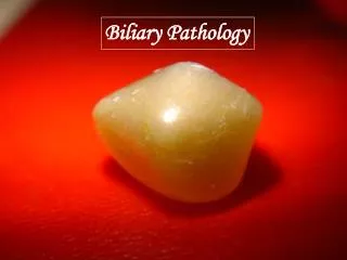
Gallstones Types: • Mixed stones (70%) – cholesterol and calcium • Pure cholesterol stones (10%) • Pigment stones – brown/black (10%) Gallstones formation: • Supersaturation of bile • Concentration of bile in the gallbladder • Crystal nucleation • Gallbladder dysmotility
Natural history of gallstones • Vast majority are asymptomatic – incidentale finding • Biliary colic – temporary obstruction of the cystic duct or gallbladder neck • 1% of patients with asymptomatic stones develop complications before onset of symptoms - prophylactic cholecystectomy is not warranted • High risk patients: • Hemolytic anemias • Porcelain gallbladder • Large (>2.5 cm) stones • Long common channels of bile and pancreatic duct • Bariatric surgery (sleeve, bypass) • Immunocompromised patients
Non-operative treatment of cholelitiasis • Generally unsuccessfull and rarely used!!! • Oral dissolution • Contact dissolution • Shock-wave lithotripsy • Up to 50% recurrence rate
Acute calculus cholecystitis • Pathogenesis: unresolved cystic duct obstruction • Inflammation, edema, subserosalhemmorhage • Infection of stagnant bile pool • Can progress to ischemia and necrosis (gangrenous cholecystitis) • Presentation: • Fever • RUQ pain • Tenderness to palpation • Laboratory finding: leukocytosis, mild elevation of bilirubin, transaminases, alk-phos.
diagnosis 1. Transabdominal US • sensitive, inexpensive and reliable • Sensitivity – 85%, specificity – 95% • Gallstones, gallbladder wall thickening, pericholic fluid, sonographicmurphy’s sign 2. Hida scan • Atypical cases • Cystic duct obstruction 3. CT scan - Less sensitive then US
Treatment • NPO • IV fluids • IV antibiotics (broad-spectrum) • Pain control • Cholecystectomy (open/lap.) • Percutaneouscholecystostomy
Chronic cholecystitis • Inflammation and scarring of the gallbladder neck and cystic duct • Pathogenesis: recurrent biliary colic which cause temporary cystic obstruction and do not cause acute cholecystitis • Presentation: recurrent biliary colic (usually after fatty meals), nausea, vomiting • RUQ/epigatric pain radiating to the scapula, usually resolves within few hours • Symptomatic cholelithiasis – indication for chlecystectomy
Diagnosis • History • Transabdominal US – stones, sludge Treatment • Elective cholecystectomy • Curative in > 90% of patients
choledocholithiasis • Primary common duct stones • De novo in the bile duct • Usually brown pigment stones • More common in Asian population • Associated with bacterial bile duct infection 2. Secondary common duct stones • Arising from the gallbladder • Most common bile duct stones in the USA • Retained common duct stones – found within 2 years of cholecystectomy
Presentation • 80-90% of common duct stones remain clinically silent • Routine cholangiography – 10% choledocholithiasis • Selective cholangiography (pain, abnormal liver function test) – 1-2% of patients will present with retained stones • Symptoms: • Biliary colic • Obstructive juandice • Ascending cholangitis (fever, pain, juandice)
Diagnosis • Hepatic function panel abnormalities • Leukocytosis • US – choledocholithiasis, biliaryductal dilatation, gallstones • Bile duct dilatation (>8 mm) in the presence of biliary colic, juandice or gall stones is suggestve of choledocholithiasis • ERCP • highly sensitive and specific • Usually therapeutic • Sphincterotomy, balloon stone extraction • Complication rate – 5-8% • MRCP - highly sensitive and specific - Does not provide therapeutic solution
treatment • ERCP • Sphincterotomy and stone extraction • Reasons for endoscopic failure: large stones, multiple stones, intrahepatic stones, altered anatomy, duodenal diverticula, impacted stones • Does not eliminate the risk of recurrent biliary stone disease (up to 50% recurrence) • Common bile duct exploration (lap./open) • Intraoperativecholangiogram • Trans-cystic/common duct incision
Ascending cholangitis • Acute ascending bacterial infection of the biliary tree cause by obstruction • Obstruction: stones, malignancy • Presentation: Charcot’s triad (fever, RUQ pain, jaundice), Reynold’s pentad (+hypotension, altered mental status)
Diagnosis • Tachycardia, shock symptoms • Laboratory test: leukocytosis, abnormal liver panel • US – dilatation of the biliary tree • CT – site of obstruction • ERCP/PTC – diagnostic as well as therapeutic
Treatment • NPO • IV fluids • IV antibiotics • Most patients respond to medical therapy • Emergent decompression of the biliary tree (ERCP/PTC)