Download
1 / 5
50 likes | 56 Views
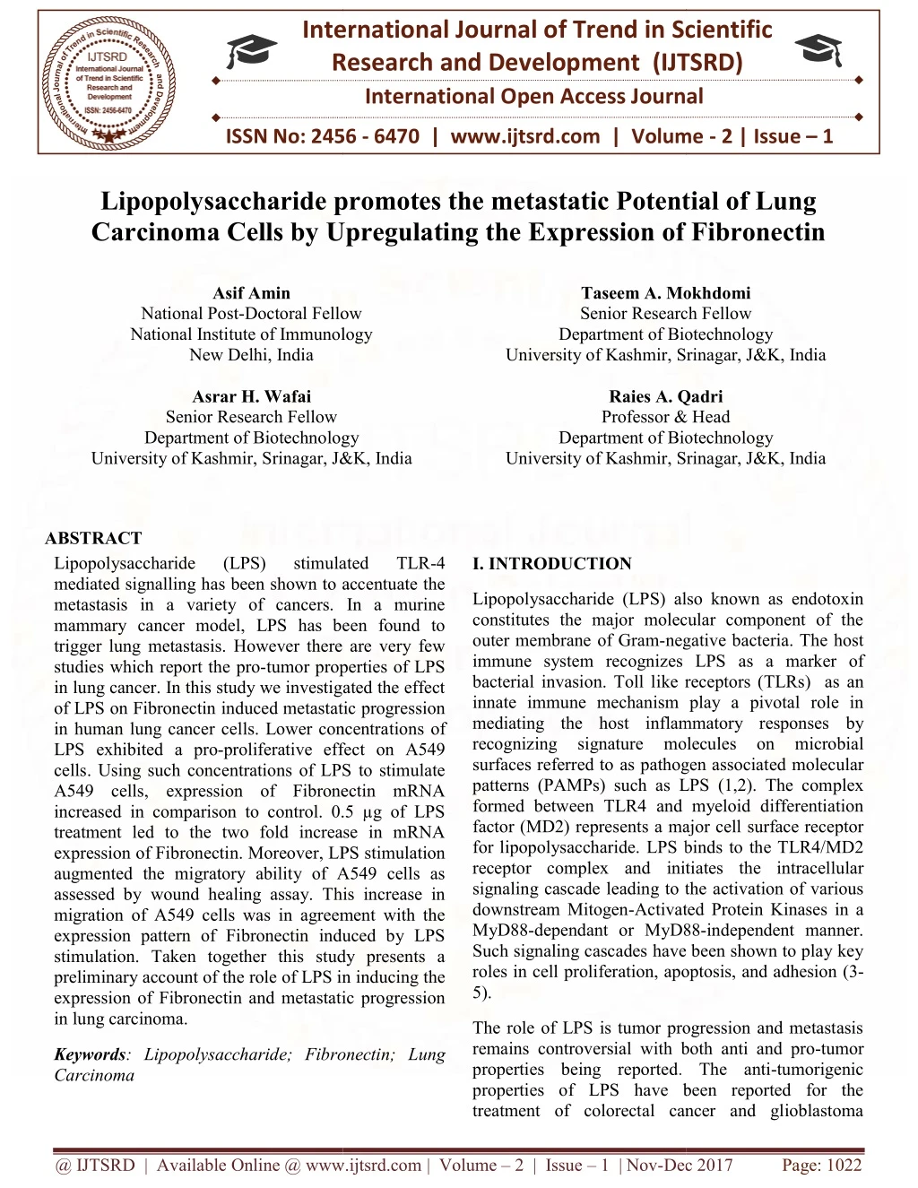
Lipopolysaccharide LPS stimulated TLR 4 mediated signalling has been shown to accentuate the metastasis in a variety of cancers. In a murine mammary cancer model, LPS has been found to trigger lung metastasis. However there are very few studies which report the pro tumor properties of LPS in lung cancer. In this study we investigated the effect of LPS on Fibronectin induced metastatic progression in human lung cancer cells. Lower concentrations of LPS exhibited a pro proliferative effect on A549 cells. Using such concentrations of LPS to stimulate A549 cells, expression of Fibronectin mRNA increased in comparison to control. 0.5 u00b5-g of LPS treatment led to the two fold increase in mRNA expression of Fibronectin. Moreover, LPS stimulation augmented the migratory ability of A549 cells as assessed by wound healing assay. This increase in migration of A549 cells was in agreement with the expression pattern of Fibronectin induced by LPS stimulation. Taken together this study presents a preliminary account of the role of LPS in inducing the expression of Fibronectin and metastatic progression in lung carcinoma. Asif Amin | Taseem A. Mokhdomi | Asrar H. Wafai | Raies A. Qadri "Lipopolysaccharide promotes the metastatic Potential of Lung Carcinoma Cells by Upregulating the Expression of Fibronectin" Published in International Journal of Trend in Scientific Research and Development (ijtsrd), ISSN: 2456-6470, Volume-2 | Issue-1 , December 2017, URL: https://www.ijtsrd.com/papers/ijtsrd7164.pdf Paper URL: http://www.ijtsrd.com/biological-science/cell-biology/7164/lipopolysaccharide-promotes-the-metastatic-potential-of-lung-carcinoma-cells-by-upregulating-the-expression-of-fibronectin/asif-amin<br>

E N D
International Research Research and Development (IJTSRD) International Open Access Journal International Open Access Journal International Journal of Trend in Scientific Scientific (IJTSRD) ISSN No: 2456 - 6470 | www.ijtsrd.com | Volume Lipopolysaccharide promotes the metastatic Potential of Lung Lipopolysaccharide promotes the metastatic Potential of Lung ISSN No: 2456 | www.ijtsrd.com | Volume - 2 | Issue – 1 Lipopolysaccharide promotes the metastatic Potential of Lung Carcinoma Cells by Upregulating the Expression of Fibronectin Carcinoma Cells by Upregulating the Expression of Fibronectin Carcinoma Cells by Upregulating the Expression of Fibronectin Asif Amin Doctoral Fellow Taseem A. Mokhdomi Senior Research Department of Biotechnology University of Kashmir, Srinagar, J&K, Taseem A. Mokhdomi Research Fellow Department of Biotechnology National Post-Doctoral Fellow National Institute of Immunology New Delhi, India National Institute of Immunology Srinagar, J&K, India Asrar H. Wafai Senior Research Fellow Department of Biotechnology University of Kashmir, Srinagar, J&K, Raies A. Qadri Professor & Head Department of Biotechnology University of Kashmir, Srinagar, J&K, India Raies A. Qadri Professor & Head artment of Biotechnology Srinagar, J&K, India Department of Biotechnology Srinagar, J&K, India ABSTRACT Lipopolysaccharide mediated signalling has been shown to accentuate the metastasis in a variety of cancers. In a murine mammary cancer model, LPS has been found to trigger lung metastasis. However there are very few studies which report the pro-tumor properties of LPS in lung cancer. In this study we investigated the effect of LPS on Fibronectin induced metastatic progression in human lung cancer cells. Lower concentra LPS exhibited a pro-proliferative effect on A549 cells. Using such concentrations of LPS to stimulate A549 cells, expression of Fibronectin mRNA increased in comparison to control. 0.5 µg of LPS treatment led to the two fold increase in mRNA expression of Fibronectin. Moreover, LPS stimulation augmented the migratory ability of A549 cells as assessed by wound healing assay. This increase in migration of A549 cells was in agreement with the expression pattern of Fibronectin induced by LPS stimulation. Taken together this study presents a preliminary account of the role of LPS in inducing the expression of Fibronectin and metastatic progression in lung carcinoma. Lipopolysaccharide mediated signalling has been shown to accentuate the metastasis in a variety of cancers. In a murine mammary cancer model, LPS has been found to igger lung metastasis. However there are very few (LPS) (LPS) stimulated stimulated TLR TLR-4 I. INTRODUCTION Lipopolysaccharide (LPS) also known as endotoxin constitutes the major molecular component of the negative bacteria. The host immune system recognizes LPS as a marker of Toll like receptors (TLRs) as an innate immune mechanism play a pivotal role in the host inflammatory responses by signature molecules on microbial as pathogen associated molecular Lipopolysaccharide (LPS) also known as endotoxin constitutes the major molecular component of the outer membrane of Gram-negative bacteria. immune system recognizes LPS bacterial invasion. Toll like receptors ( innate immune mechanism play a mediating the host inflammatory responses recognizing signature molecules surfaces referred to as pathogen associat patterns (PAMPs) such as LPS formed between TLR4 and myeloid differentiation factor (MD2) represents a major cell surface receptor for lipopolysaccharide. LPS bind receptor complex and initiates the intracellular signaling cascade leading to the activation of various downstream Mitogen-Activated Protein Kinases MyD88-dependant or MyD88 Such signaling cascades have been shown to play key roles in cell proliferation, apoptosis, and adhesion 5). tumor properties of LPS in lung cancer. In this study we investigated the effect of LPS on Fibronectin induced metastatic progression in human lung cancer cells. Lower concentrations of proliferative effect on A549 cells. Using such concentrations of LPS to stimulate A549 cells, expression of Fibronectin mRNA increased in comparison to control. 0.5 µg of LPS treatment led to the two fold increase in mRNA ssion of Fibronectin. Moreover, LPS stimulation augmented the migratory ability of A549 cells as assessed by wound healing assay. This increase in migration of A549 cells was in agreement with the expression pattern of Fibronectin induced by LPS n. Taken together this study presents a preliminary account of the role of LPS in inducing the expression of Fibronectin and metastatic progression LPS (1,2). The complex formed between TLR4 and myeloid differentiation a major cell surface receptor LPS binds to the TLR4/MD2 initiates the intracellular leading to the activation of various Activated Protein Kinases in a D88-independent manner. have been shown to play key ation, apoptosis, and adhesion (3- The role of LPS is tumor progression and metastasis remains controversial with both anti and pro properties being reported. properties of LPS have been reported for the treatment of colorectal cancer and glioblastoma treatment of colorectal cancer and glioblastoma The role of LPS is tumor progression and metastasis with both anti and pro-tumor Keywords: Lipopolysaccharide; Fibronectin; Lung Carcinoma Lipopolysaccharide; Fibronectin; Lung . The anti-tumorigenic been reported for the @ IJTSRD | Available Online @ www.ijtsrd.com @ IJTSRD | Available Online @ www.ijtsrd.com | Volume – 2 | Issue – 1 | Nov-Dec 2017 Dec 2017 Page: 1022
International Journal of Trend in Scientific Research and Development (IJTSRD) ISSN: 2456-6470 multiforme. An increasing body of evidence also points at the role of LPS in tumor progression. Stimulation of TLR4 by LPS has been found to trigger increased lung metastasis in a murine mammary cancer model of metastatic disease. This effect was ascribed to an increased proliferation and inhibition of apoptosis of tumor cells as well as an tumor cell invasion and migration along with increased angiogenesis (6,7). Besides this study, TLR4 has been implicated in tumor cell invasion, survival, and metastasis in many cancer types (8). A recent study demonstrated that LPS can increase the migration ability of human cell esophageal cancer HKESC-2 cells via signaling through TLR4 (9). Similarly in breast cancer TLR4 activation by LPs has been shown to promote the αvβ3-mediated adhesion and invasiveness (10). D. Reverse transcriptase polymerase chain reaction RNA was isolated using TRIzol reagent according to manufacturer’s instructions. Briefly 1 ml of TRIzol was added to the cells grown in 30 mm culture dish followed by homogenization using a pipette. Thereafter 1ml of sterile water was added and the mixture incubated at room temperature for 5-15 min. The mixture was centrifuged at 12,000 g for 15 min and supernatant collected, to which 70% isopropanol was added. The supernatant was further centrifuged at 12,000 g for 10 min to obtain the RNA pellet which was washed two times with 75% ethanol. The integrity of RNA was checked on 1% agarose gel. In succession to RNA isolation and subsequent quantification, RNA was reverse transcribed to cDNA using reverse transcriptase and oligo-dT primers in a final volume of 20 μl. II.Materials and Methods The primers used for evaluating the expression status of Fibronectin are as: A. Chemicals and reagents DMEM and FBS were procured from Gibco, USA. Penstrip was obtained from Invitrogen, USA. LPS (Escherichia coli 0111:B4), DMSO and MTT were purchased from Sigma Chemical Co. (St. Louis, MO, USA). LPS was prepared as a stock solution of 1 mg/ml and was diluted to various concentrations with serum free medium when used. Forward 5…AGTCAGCCTCTGGTTCAGAC...3 primer Reverse 5…CTTCAGGTCAGTTGGTGCAG...3 primer PCR products were analyzed on 1–2% agarose gels and the ratio of TLR4 to β-actin served as the level of mRNA expression. B. Cell culture Human lung carcinoma cell line, A549 was provided by Dr. Ayub Qadri, Immunology. A549 Cells were cultured in DMEM, supplemented with 10% fetal bovine serum, and antibiotics (1% penstrip) at 37 °C in 5% CO2 humidified incubator. The medium was changed regularly and the cells were sub-cultured when confluent. National Institute of E. Real Time PCR Real-time PCR was performed to detect the gene expression of Fibronectin in A549 cells under LPS stimulation. The 10 µl reaction mixture contained 5.5 µl nuclease-free water, 1.0 ml cDNA (1 mg/ml), 0.5 µl (10 mM) each primer and 2.5 µl Light Cycler 480 SYBR Green Master (Roche, Germany). The amplification was carried at 94 °C for 2 min incubation and 40 cycles of 94 °C for 15 s, 56 °C for 15 s, 72 °C for 20 s, followed by final extension of 72 °C for 2 min in LightCycler 480 II (Roche, Germany). Relative quantification was done using 2- ΔΔcp method (11). C. MTT Cell Proliferation Assay Cells were seeded into the wells of a 96 well plate and allowed to grow overnight. LPS was added to cells at various concentrations (0.1, 0.5, 1, 1.5, 2 and 2.5 μg) for 24 h. Following treatment, 100 µl of MTT solution was added to each well followed by 2 h incubation at 37°C in dark. Then the MTT solution was removed and 100 µl of DMSO was added to cells. The quantification was done by measuring the absorbance at 590 nm (background 650 nm) using micro plate reader (Biotek, USA) and the percentage proliferation was calculated. F. Cell migration Assay Equal number of A549 cells was seeded in each well of 24-well plate and allowed to grow to sub- confluency. Then the scratch was created using a 100 μl pipette tip followed by washings with PBS to @ IJTSRD | Available Online @ www.ijtsrd.com | Volume – 2 | Issue – 1 | Nov-Dec 2017 Page: 1023
International Journal of Trend in Scientific International Journal of Trend in Scientific Research and Development (IJTSRD) ISSN: 2456 Research and Development (IJTSRD) ISSN: 2456-6470 remove the floating cells. This was followed by treatment with sub-toxic concentrations of LPS for 24 h. Photographs were taken before (0 h) and after treatment at 24 h. Wound healing was calculated as the percentage of the initial wound before treatment to the total wound closure at 24 h after treatment. The assay was performed three independent times. assay was performed three independent times. remove the floating cells. This was followed by toxic concentrations of LPS for 24 h. Photographs were taken before (0 h) and after μg) of LPS for 24 h. Following treatment, RNA was isolated from A549 cells using trizol reagent 2). h. Following treatment, RNA was isolated from A549 cells using trizol reagent (Figure as calculated as the percentage of the initial wound before treatment to the total wound closure at 24 h after treatment. The III.RESULTS A. Determination of Sub-lethal doses of LPS doses of LPS Exposure to bacterial LPS has been shown to induce apoptotic cell death. In order to determine the lethal doses of LPS, A549 cells were treated with different concentrations of LPS. It was observed that LPS did not result in appreciable inhibition of c proliferation up to 1μg of LPS. However exceeding 1μg had a significant impact on the proliferation of A549 cells. Therefore we only used sub-lethal concentrations of LPS for further (Figure 1). Exposure to bacterial LPS has been shown to induce order to determine the Sub- were treated with Figure 2. RNA isolation from treated cells . RNA isolation from control and LPS different concentrations of LPS. It was observed that LPS did not result in appreciable inhibition of cell g of LPS. However the doses g had a significant impact on the proliferation of A549 cells. Therefore we only used lethal concentrations of LPS for further assays The extracted RNA was reverse transcribed to cDNA using oligo dT primers. To evaluate the effect of LPS treatment on the expression of Fibronectin at mRNA level, cDNA obtained was amplified using specific primers. It was observed that the treatment with LPS led to the increase in the expression of Fibronectin g concentration (Figure 3 A). However at 0.1 µg there was decrease in the gene expression of Fibronectin as compared to untreated For further quantitative analysis, real-time PCR was carried out and the results obtained were in consonance with those obtained by semi-quantitative Real-Time PCR analysis revealed that the Fibronectin gene expression gets upregulated 2 fold using 0.6 µg LPS in comparision The extracted RNA was reverse transcribed to cDNA using oligo dT primers. To evaluate the effect of LPS treatment on the expression of Fibronectin at mRNA level, cDNA obtained was amplified using specific primers. It was observed that the treatment with LP led to the increase in the expression of particularly at 0.6 µg concentration (Figure 3 A However at 0.1 µg there was decrease in the gene expression of Fibronectin as compared to untreated control. For further quantitative analysis, real PCR was carried out and the results obtained were in consonance with those obtained by semi analysis (Figure 3B). Real revealed that the Fibronectin gene expression gets upregulated 2 fold using 0.6 µg LPS in comparision to untreated control. Figure 1. Evaluation of sub-lethal dos A549 cells by MTT assay. A549 cells were treated with the indicated concentrations of LPS for 24 h. After 24 h, cytotoxicity was determined by MTT assay. Data represented as mean ± SD of results from three independent experiments. doses of LPS on A549 cells by MTT assay. A549 cells were treated with the indicated concentrations of LPS for 24 h. After 24 h, cytotoxicity was determined by MTT assay. Data represented as mean ± SD of results from B. Treatment with LPS increases the expression of Fibronectin in A549 cells with LPS increases the expression of Fibronectin has been shown to promote metastatic behavior of many tumors including those of lung. Therefore to assess the effect of LPS on the expression of Fibronectin, A549 cells to various sub-lethal concentrations (0.1, 0.5 and 1 Fibronectin has been shown to promote metastatic behavior of many tumors including those of lung. to assess the effect of LPS on the cells were exposed (0.1, 0.5 and 1 @ IJTSRD | Available Online @ www.ijtsrd.com @ IJTSRD | Available Online @ www.ijtsrd.com | Volume – 2 | Issue – 1 | Nov-Dec 2017 Dec 2017 Page: 1024
International Journal of Trend in Scientific International Journal of Trend in Scientific Research and Development (IJTSRD) ISSN: 2456 Research and Development (IJTSRD) ISSN: 2456-6470 C. Treatment with LPS accentuates the migration of A549 cells Treatment with LPS accentuates the migration Migration and invasion represent the processes Fibronectin has been shown to enhance the migration and invasiveness of lung carcinoma cells. To evaluate the effect of LPS on the migration of A549 cells, a wound healing assay was A549 cells were grown to confluency, wounded and then treated . It was observed that LPS stimulation led to the healing of wound with best Migration and invasion represent the processes crucial to metastasis. Fibronectin has been shown to enhance the migration and invasiveness of lung carcinoma cells. To evaluate the effect of LPS migration of A549 cells, a wound healing assay was carried out. A549 cells were grown to confluency, wounded and then treated concentration of LPS. It was observed stimulation led to the healing of w concentration being 0.5 μg. In terms of percentage wound healing, while treatment with 1 μg and 0.5 led to the healing of wound to about 86% and 95% respectively while as 0.1 μg was not much effective when compared with the control when compared with the control (Figure 4). with with indicated indicated . In terms of percentage healing, while treatment with 1 μg and 0.5 μg led to the healing of wound to about 86% and 95% respectively while as 0.1 μg was not much effective Figure 3. Gene expression analysis of Fibronectin LPS stimulated A549 cells by (A) Semi quantita reverse transcription PCR (B) Real Time PCR represented as mean ± SD of results from three independent experiments. Fibronectin in by (A) Semi quantitative reverse transcription PCR (B) Real Time PCR. Data represented as mean ± SD of results from three Figure 4. Effect of LPS on the migration of A549 cells. A549 cells were grown to sub and treated with indicated concentrations of LPS or left untreated 24h. (B) The data calculated from three independent experiments and represented as mean ± SD. 24h. (B) The data calculated from three independent experiments and represented as mean ± SD. 24h. (B) The data calculated from three independent experiments and represented as mean ± SD. Effect of LPS on the migration of A549 cells. A549 cells were grown to sub and treated with indicated concentrations of LPS or left untreated (control). (A) Wound healing observed Effect of LPS on the migration of A549 cells. A549 cells were grown to sub-confluency, wounded (control). (A) Wound healing observed after promoting the tumor cell migration and invasion (12). Of particular importance is its role in epithelial- mesenchymal transition which is a cellular program indispensible to cancer metastasis (13). Pathogen promoting the tumor cell migration and invasion (12). Of particular importance is its role in mesenchymal transition which is a cellular program indispensible to cancer metastasis associated molecular patterns such as LPS have associated molecular patterns such as LPS have IV.DISCUSSION Fibronectin is an extracellular matrix glycoprotein that has been shown to promote metastatic process in a wide variety of cancer types. It has been implicated in It has been implicated in Fibronectin is an extracellular matrix glycoprotein that has been shown to promote metastatic process in a wide @ IJTSRD | Available Online @ www.ijtsrd.com @ IJTSRD | Available Online @ www.ijtsrd.com | Volume – 2 | Issue – 1 | Nov-Dec 2017 Dec 2017 Page: 1025
profound consequences on tumor growth as they have been found to promote cancer progression and anti- apoptotic activity (6-8). In a recent study NADPH oxidase 1-dependent ROS was demonstrated to be crucial for TLR4 signaling to promote tumor metastasis of non-small cell lung cancer (14). In this way, we therefore designed this study to determine the action of LPS on Fibronectin induced metastatic progression in lung carcinoma cells. A549 cells were stimulated with sub-lethal doses of LPS. At such concentrations there was an increase in the proliferation of these cells. Furthermore treatment with such concentrations led to increase in the expression of Fibronectin at mRNA level as was assessed by semi-quantitative reverse transcription PCR as well as quantitative real time PCR. To further assess the effect of LPS on the migration, A549 cells were treated with sub-lethal concentrations of LPS. It was observed that the treatment with LPS increased the migratory ability of A549 cells as was depicted by significant closure of the wound. The finding was in coherence with the upregulation of Fibronectin by LPS treatment. Thus it can be argued that LPS accentuates the metastatic process in lung cancer cells by upregulating the expression of Fibronectin. The present study presents a preliminary account of effect of LPS on Fibronectin induced metastatic progression in lung carcinoma and thus warrants an exhaustive study to discern the mechanistic action of LPS. International Journal of Trend in Scientific Research and Development (IJTSRD) ISSN: 2456-6470 of induced tumour growth in a murine model of metastatic disease. British Journal of Cancer. 81: 1311–7. endotoxin/lipopolysaccharide in surgically 7)[7] Harmey JH, Bucana CD, Lu W, Byrne AM, McDonnell S, Lynch Lipopolysaccharide-induced metastatic growth is associated with increased angiogenesis, vascular permeability and tumor cell invasion. International Journal of Cancer.101: 415–22. C, et al. (2002) 8)[8] Yang H, Wang B, Wang T, Xu L, He C, Wen H, et al. (2014) Toll-like receptor 4 prompts human breast cancer cells lipopolysaccharide stimulation and is overexpressed in patients with lymph node metastasis. PLoS ONE .9: e109980 invasiveness via 9)[9] Rousseau MC, Hsu RY, Spicer JD, Mcdonald B, Chan CH, Perera RM, et al. (2013) Lipopolysaccharide-induced toll-like receptor 4 signaling enhances the migratory ability of human esophageal cancer cells in a selectindependent manner. Surgery.154: 69-77. 10)[10] Liao SJ, Zhou YH, Yuan Y, Li D, Wu FH, et al. (2012) Triggering of Toll-like receptor 4 on metastatic breast cancer cells promotes avb3- mediated adhesion and invasive migration. Breast Cancer Research and Treatment. 133: 853–63. 11)[11] Livak KJ, Schmittgen TD (2001) Analysis of relative gene expression data using real- time quantitative PCR and the 2-ΔΔcp Method. Methods. 25: 402–408 References 1)[1] Janeway CA Jr, Medzhitov R. (2002) Innate immune recognition. Immunolgy. 20: 197–216. Annual Reviews of 12)[12] Ou, J., Peng, Y., Deng, J., Miao, H., Zhou, J., Zha, L., Liang, H. (2014). Endothelial cell-derived fibronectin extra domain A promotes colorectal cancer metastasis via mesenchymal transition. Carcinogenesis. 35: 1661– 1670 2)[2] Akira S, Uematsu S, Takeuchi O. (2006) Pathogen recognition and innate immunity. Cell 124: 783–801. inducing epithelial– 3)[3] Kim HM, Park BS, Kim JI, Kim SE, Lee J, Oh SC, et al (2007) Crystal structure of the TLR4-MD- 2 complex with bound endotoxin antagonist Eritoran. Cell 130: 906–17. 13)[13] Amin, A., Chikan, N. A., Mokhdomi, T. A., Bukhari, S., Koul, A. M., Shah, B. A., & Qadri, R. A. (2016). Irigenin, a novel lead from Western Himalayan chemiome inhibits Fibronectin-Extra Domain A induced metastasis in Lung cancer cells. Scientific Reports. 6. 4)[4] Park BS, Song DH, Kim HM, Choi BS, Lee H, Lee JO. (2009) The lipopolysaccharide recognition by the TLR4-MD-2 complex. Nature 458: 1191–5. structural basis of 14)[14] Liu X, Pei C, Yan S, Liu G, Liu G, Chen W et al. (2015) NADPH oxidase 1-dependent ROS is crucial for TLR4 signaling to promote tumor metastasis of non-small cell lung cancer. Tumor Biology. 6: 1493–1502. 5)[5] Lu YC, Yeh WC, Ohashi PS. (2008). LPS/TLR4 signal transduction pathway. Cytokine 42: 145–51. 6)[6] Pidgeon GP, Harmey JH, Kay E, Da Costa M, Redmond HP, Bouchier-Hayes DJ. (1999) The role @ IJTSRD | Available Online @ www.ijtsrd.com | Volume – 2 | Issue – 1 | Nov-Dec 2017 Page: 1026