Download

1 / 38
400 likes | 860 Views
Metastatic Carcinoma of Unknown Primary: A Diagnostic Dilemma. Lalan S. Wilfong, MD Texas Oncology, PA February 23, 2006. Overview. Definition Epidemiology Biology Diagnostic Work-up Clinical Radiology Pathology Specific Clinical Syndromes Treatment. Definition.

E N D
Metastatic Carcinoma of Unknown Primary: A Diagnostic Dilemma Lalan S. Wilfong, MD Texas Oncology, PA February 23, 2006
Overview • Definition • Epidemiology • Biology • Diagnostic Work-up • Clinical • Radiology • Pathology • Specific Clinical Syndromes • Treatment
Definition • Metastatic Cancer of Unknown Primary • Biopsy confirmed malignancy • for which the site of origin cannot be identified by routine workup • Primary lesion can be identified in only 30-80% of cases at autopsy • Hypotheses • primary tumor has involuted and is not detectable • Malignant phenotype favors metastases over primary tumor growth
Epidemiology • Accounts for 5-10% of cancer diagnoses • Median survival of approximately 6-12 months despite therapy • However, certain subgroups are potentially curable • Factors relating to overall survival • age • sex • lymph node vs visceral mets
Biology • Heterogeneous group of malignancies, but share common features • presence of early metastases • maybe useful model to understand early tumor invasion and distant spread • 30% have 3 or more organs involved compared to only 15% of patients with known primary • Unusual metastatic pattern involving kidneys, adrenal, skin and heart
Aneuploidy chromosome instability found in 70-90% of tumors usually implies worse prognosis Chromosomal Abnormalities loss of short arm of chromosome 1 13/30 patients studied Overexpress Oncogenes c-myc bcl-2 her 2 neu Inactivated tumor suppresser genes p53 Microvessel Density marker of angiogenesis worse survival Biology
Clinical Work-Up • Natural inclination is to perform extensive search for a primary • absence of primary generates anxiety • used to predicting tumor behavior and survival based on primary tumors • therapy usually based on primary tumor pathology • Typical evaluation costs between $4500 and $18,000 per patient • Total annual US costs roughly 1.5 billion dollars
History Complete ROS PMH previous moles? Biopsies? SH smoking asbestos HIV FH -- clustering of cancers can lead to syndromes Physical Thorough skin evaluation Oral and nasal cavities Lymph nodes Breast Rectal Pelvic/Genital Prostate H&P **** Important Step ****
Laboratory • Basic CBC, CMP • Recommended tumor markers • Men • PSA • bHCG • aFP • Women - none • Other markers not recommended • poor sensitivity and specificity • all can be elevated in multiple tumor types
Radiology • Recommended • CXR +/- Chest CT scan • Abdominal/Pelvic CT scan • Mammogram in women • MD Anderson experience • Primary site identified in 20% • 1/3 of these based on unique histology • No difference in survival between patients in whom a primary site identified and those whose primary remained occult
Pet Scans • Positron Emission Tomography • Utilizes [18F] Fluorodeoxyglucose (FDG) • radio-isotope of glucose • Warburg effect -- neoplastic cells undergo accelerated glycolysis • FDG concentrates in neoplastic cells to localize tumors • Theoretically could localize primary sites • Limited studies available on this topic
PET • Meta-analysis by Delgado-Bolton, et al published in The Journal of Nuclear Medicine 2003; 44:1301-1314 • 15 studies selected evaluating 302 patients • Identified primary tumor site in 129 patients • Sensitivity -- 0.87 (0.81-0.92) • Specificity -- 0.71 (0.64-0.78) • General use hindered by • lack of prospective studies • cost-effectiveness hasn’t been assessed
Primary sites Localization of Tumor
Pathology • Heterogeneous collection of tumor types • Includes • Carcinomas • Poorly differentiated malignancies • Sophisticated pathologic evaluation • Identify certain histologies • Allow appropriate therapy • Techniques • Light microscopy • Immunohistochemical staining • Electron microscopy • Molecular genetics
Squamous Carcinoma 5% PDC, PDA 30% Adenocarcinoma 60% PDMN 5% Melanoma, Sarcoma, Other 1% Lymphoma, Melanoma Sarcoma 3% PDC, PDA 1% No specific Subgroup 1% Specific Carcinoma 1% PDC, PDA 26% Specific Subgroup 4% Specific Subgroup 6% No specific Subgroup 54% Lymphoma 3% Cancer of an Unknown Primary Light Microscopical Diagnosis Specialized Pathological Study of Specific Clinical Features
Epithelial origin cytokeratins Melanoma PS100 HMB45 Germ Cell Tumor aFP bHCG PLAP Neuroendocrine Chromogranin synaptophysin Lymphoma Cd45 Cd10 Cd3 Thyroid Thyroglobulin Prostate PSA Sarcoma AML CD31 CD34 Immunohistochemistry
Ck7- Ck20+ Ck7+ Ck20+ Ck7+ Ck20- Ck7- Ck20- Broncho-pulmonary -TTF1 Breast -EMA, GCDFP, ER/PR Nonmucinous Ovarian -CA-125 Thyroid -TTF1 Cholangiocellular -CEA, Cd10 Hepatocellular -aFP Renal Cell -VIM Prostate -PSA Urothelial Pancreatic -CEA Gastric -CEA Mucinous Ovarian Colorectal -CEA Adenocarcinoma Ck7, Ck20 Clinical Signs
Molecular Genetics • Chromosomal evaluation • Well documented usefulness in hematologic malignancies • Techniques • Classical • Southern blot • FISH • PCR
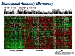
Microarray • Uses cDNA technology • Allows thousands of genes to be analyzed simultaneously • Provides organ specific genetic profile • Two investigators have correctly identified both • Primary site • Metastatic disease origin
Specific Clinical Syndromes • After complete pathologic review evaluating • Treatable diagnoses such as lymphoma • Found primary sites • Clinical syndromes can be identified • Important to recognize these syndromes • Can be potentially treatable or even curable • Based on • Histology of tumor • Location • Gender
Peritoneal Carcinomatosis in Women • Adenocarcinoma • Malignant ascites • Extensive peritoneal involvement • Most characteristic of ovarian cancer • Used to be classified as MCUP • Now classified as ovarian • Cell of origin unclear • Germinal epithelium of ovary and mesothelium of the peritoneum have the same embryologic origin • Retains multipotentiality
Peritoneal Carcinomatosis • Histology is a serous carcinoma • Ovarian primary not detectable • Can occur in women s/p oophorectomy • Small deposits of tumor can be seen on ovary surfaces • Some women have BRCA 1 mutations • Treatment similar to ovarian cancer • Surgical debulking • Followed by systemic chemotherapy • Survival • Similar to ovarian cancer at equivalent stage • Median survival 11-24 months • Five-year survival of 15-20%
Metastatic Carcinoma in Axillary Lymph Nodes in Women • Unilateral axillary lymph nodes • Most suggestive of breast primary • Careful breast evaluation • Breast exam • Mammogram • Detects primary in 25-50% • Ultrasound • MRI • One small study primary identified in 86% of 22 cases
Axillary Lymph Nodes -- Treatment • Treated like node positive breast primary • If breast primary not found on imaging • Local treatment is controversial • Historically mastectomy was done • Careful pathologic review failed to reveal a breast tumor in 33-47% of cases • Breast conservation therapy evaluated to limited extent • Axillary node dissection + breast radiation • Survival and local recurrence rates similar • Chemotherapy • Treated like node positive breast tumors • No prospective studies validate this approach • Hormonal therapy if ER+/PR+ • Prognosis based on number of positive nodes
Squamous Cell Cancer in Cervical Lymph Nodes • Presentation accounts for 1-2% of all head and neck malignancies • Lung and esophagus can present in similar fashion • Lymph nodes usually in low neck • Work-up • CT of head and neck • Panendoscopy – laryngoscopy, bronchoscopy, and esophagoscopy • Also included blind biopsies of common primary sites • Ipsilateral tonsillectomy can harbor occult primary in 10-25% of cases • Primary site still not identified in 2/3 of cases
Cervical Lymph Nodes -- Treatment • Typical approach • Neck dissection • Followed by radiation therapy • Controversy exists • Either treatment modality alone • Extent of radiation • Bilateral neck and total mucosal has high morbidity • Localized radiation to ipsilateral neck alone • Retrospective studies suggest more aggressive approach improves local control and survival • Prognosis depends on extent on lymph node involvement • Long term local control 50-75% of patients • Five-year survival 40-60%
Squamous Cell Cancer in Inguinal Lymph Nodes • Likely primary sites • Anus • Cervix, vulva or vagina in women • Lower extremities • Work-up • Lower extremity exam • Anoscopy • Genital/pelvic exam • Treatment if no primary found • Surgery +/- radiation therapy • Long term survival of 25%
Men with Possible Prostate Cancer • Older men • Predominant bony metastases – blastic • Work-up • Serum PSA • IHC of tumor for PSA • Treatment • Hormonal therapy • Some advocate even in setting of negative PSA in men with osteoblastic bone metastases
Neuroendocrine Carcinoma • Heterogeneous Group • Three identifiable subsets based on histology • Typical carcinoid or pancreatic islet cell tumors • Small cell carcinoma • Poorly differentiated carcinoma that has neuroendocrine features identified only by electron microscopy or IHC
Typical carcinoid • Often have metastatic disease to the liver • May or may not have clinical evidence of hormone production • Typically indolent tumors and progress slowly • Treatment • Chemotherapy has limited efficacy • Surgery if isolated metastases • Octreotide useful for symptomatic hormone production
Small Cell • Natural history similar to lung primary • Treated with platinum based chemotherapy • Rare long term survival can be achieved • Isolated metastasis have been reported • Only case reports published • Recommended treatment is similar to limited stage small cell • Radiation • chemotherapy
Poorly Differentiated Neuroendocrine Carcinoma • One series published by Hainsworth, et al • Represented a particularly chemosensitive group of patients • Reported response rate to platinum based chemotherapy of over 60% • Long term survival of 10%
Extragonadal Germ Cell Tumor • Clinical presentation consistent with metastatic germ cell tumor but lack definitive histology • Men <50 • Midline tumors (retroperitoneum, mediastinum) and/or pulmonary nodules • Duration of symptoms short or rapid tumor growth • Elevated aFP, bHCG • (i)12p on molecular genetics • Usually respond well to platinum based chemotherapy • Survival similar to primary germ cell tumor based on tumor markers and location of disease
Prognosis of MCUP • Prognosis • Median survival 6-12 months • 5-10% survival at 5 years • Poor prognostic factors • Male gender • Liver mets • Increasing number of organs involved • Performance status
Regression Tree Analysis No patients Liver = No Median Survival in months Bone = No Bone = Yes Liver = Yes Adrenal = No Adrenal = Yes Path = Neuro Path = Adeno, Squamous Pleura = No Pleura = Yes # sites <2.0 # sites >2.0 Age <61 Age >61 153 Path = Neuro, squamous 5 Path = Adeno 127 40 Adapted from Hess, et al Clin Cancer Res 1999; 5:3403-10
Treatment • Historically combination chemotherapy used • 5fu, cisplatin, adriamycin or mitomycin • Response rates 0-40% • Median survival 3-8 months • Recent combinations included taxanes • Carboplatin, paclitaxel and oral etoposide • Hainsworth et al reported • Response rate of 47% • Median survival of 13 months • Other trials not as impressive results
Newer agents • Gemcitabine and Docetaxel combination • Cisplatin refractory disease • Response rate 28% • Median survival 8 months • Molecular agents • Herceptin for Her-2-neu positive disease • VEGF inhibitors • EGFR inhibitors • Proteosome inhibitors
Conclusions • MCUP is a common heterogeneous disease • Work-up • History and Physical • Limited radiographs • Pathology • Light microscopy • IHC • Specialized techniques • Identify specific clinical syndromes • Treatment can be given