Download

1 / 14
140 likes | 278 Views
Implantation of the blastocyst in the guinea pig Allen C Enders Department of Cell Biology and Human Anatomy University of California, Davis. Implantation in the guinea pig.

E N D
Implantation of the blastocyst in the guinea pig Allen C Enders Department of Cell Biology and Human Anatomy University of California, Davis
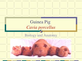
Implantation in the guinea pig The guinea pig is the common laboratory example of histricognath rodents. The blastocyst in these species has thickened abembryonictrophoblast which becomes syncytial, forming an implantation cone from which ectoplasmic processes extend through the zonapellucida. These processes effect adhesion to the uterine luminal epithelium antimesometrially. Subsequently the trophoblast penetrates the luminal epithelium and apparently draws the rest of the blastocyst into the endometrial stroma. It is thus an interstitial implantation. However, shortly after the inner cell mass has been drawn into the undecidualized endometrium, the syncytial trophoblast that is abembryonic begins to degenerate. This degeneration exposes the apical surface of the endoderm, which covers the inner cell mass (ICM) and attaches to healthy trophoblast around the ICM, to a space within the decidualizing endometrium. Later the disintegration of the adjacent luminal epithelium completes the early inversion of the yolk sac and exposes it to the uterine lumen. This inversion is earlier than in most myomorph rodents (sciuromorph rodents do not have complete inversion). Guinea pigs in our colony generally mated between 5 pm and 8 am, apparently more commonly towards the end of this time period. Penetration of the zonapellucida, attachment, and invasion of the uterine luminal epithelium usually occured after an elapsed time of 6 days. By 7 days intramural blastocysts showed some trophoblast degeneration, which was quite marked by 8 days.
2 Early blastocyst. Note the differentiation of the endoderm under the epiblast cells of the inner cell mass (ICM). The endoderm does not spread to a parietal position in this species. • 1 Section through a four-cell stage. • Twenty six hours after fertilization, three blastomeres are surrounded by a zonapellucida, whose contrast has been enhanced.
3 A blastocyst flushed at the time of attachment. Some cells of the ICM are seen above, and processes of the abembryonic syncytial trophoblast are seen below penetrating into the zonapellucida, which is barely visible. 4 Section through abembryonictrophoblast (implantation cone) of a flushed blastocyst. Note the syncytial mass with its projections into the pale zonapellucida.
5 Electron micrograph of abembryonictrophoblast. The projections from the syncytial mass extend well into the zonapellucida. 6 Abembryonictrophoblast with projections. Two areas of extrusion of syncytial trophoblast into and through the zonapellucida are seen in this micrograph.
7 Implanting blastocyst clasped within the endometrium. A few inner cell mass cells appear above, and a bit of abembryonic syncytial trophoblast is seen below. Note the intact zonapellucida. 8 Section of the same blastocyst in figure 7. The abembryonictrophoblast has sent processes through the zonapellucida, contacting the underlying uterine luminal epithelium.
9 Further section of the blastocyst seen in figure 7. The trophoblast process through the zonapellucida is also seen in this section. 10 Electron micrograph of attaching blastocyst. A tongue of trophoblast at the right has penetrated the zonapellucida and formed adhesion junctions with the underlying uterine epithelial cells.
11 Beginning of intramural invasion. The blastocyst has penetrated through the uterine epithelium, and all but a little of the area near the ICM is now within the endometrium. In this micrograph the epiblast cells are completely surrounded by endoderm. Syncytial trophoblast masses (dark) appear both above and below the ICM. 12 A largely intramural blastocyst. Note that there has been very little disruption of the uterine luminal epithelium where the blastocyst entered the endometrium.
13 Higher magnification of the blastocyst seen in the figure 12. There is trophoblast both above and below the developing ICM and endoderm. The trophoblast below shows some vacuolization, and for the first time the endometrial stromal cells are beginning to show decidualization. 14 Endometrial stroma. A gland and variably decidualized stromal fibroblasts are present in an area that is near the implanting blastocyst.
16 Higher magnification of the ICM in figure 15. Note the mitotic figure in the endoderm that surrounds the pale epiblast cells. Only the syncytial trophoblast that extends in a mesometrial direction is seen in this micrograph. 17 Intramural blastocyst at the beginning of trophoblast degeneration. The endometrium adjacent to the blastocyst is well-decidualized. The syncytial trophoblast below (antimesometrial) is beginning to show signs of degeneration. 15 Intramural blastocyst in decidualized endometrium.
18 Intramural blastocyst at beginning of inversion. The extensive degeneration of the syncytial trophoblast precedes the breakdown of the uterine luminal epithelium, which will result in the early exposure of blastocyst endoderm to the uterine luminal contents, i.e. complete yolk sac inversion. 19 Higher magnification of figure 18. The ICM appears as a ball suspended by the surrounding endoderm. The trophoblast above will eventually form a mesometrially-situated placenta. The uterine epithelium has not yet degenerated to complete the inversion of the yolk sac.
21 ICM and adjacent endoderm of the previous micrograph. Note the polarity of the endodermal cells, i.e. their apical ends face outward toward the surrounding space. There is no evidence of mesoderm formation at this stage. 20 Blastocyst in the process of yolk sac inversion. Again the ICM is suspended within the endoderm in a space created by the degeneration of the abembryonictrophoblast.
22 The mesometrial-facing area of the implantation site in figure 21. A small mass of syncytial trophoblast is in the upper left. A cluster of cytotrophoblast cells tethers the endoderm; this trophoblast will contribute to forming the placenta. 23 Decidual cells at inversion. Note that the cells on the right are well-decidualized. However the stromal tissue adjacent to the cytotrophoblast of the attaching blastocyst (in the lower left) is less decidualized and has more extracellular space.
Conclusions and interpretation Early stages of implantation in the guinea pig are difficult to find within the uterine lumen. Penetration into the endometrium by the blastocyst precedes decidualization. The uterine wall is thick and the uterine lumen is highly irregular. In addition the blastocyst appears to partially collapse, perhaps facilitating the drawing of the ICM into the endometrium by the invading syncytial trophoblast. Despite the successful invasion of the syncytial trophoblast into the endometrium and its initial expansion within the endometrial stroma, the more antimesometrial portion of this trophoblast soon begins to disintegrate. This disintegration initiates the process of yolk sac inversion. It would be extremely interesting to study the factors that lead to the selective demise of this previously invasive trophoblast. Enders AC, Schlafke S 1969 Cytological aspects of trophoblast-uterine interaction in early implantation. Am J Anat 125:1-30.