Download
1 / 50
500 likes | 575 Views
Fundamentals of the Nervous System. Three functions of the nervous system 1- sensory (afferent) input: sensory receptors that work with the NS. 2- integration: processing and interpreting of data from the sensory input 3. motor (efferent) output: a response initiated by integration.

E N D
Three functions of the nervous system 1- sensory (afferent) input: sensory receptors that work with the NS. 2- integration: processing and interpreting of data from the sensory input 3. motor (efferent) output: a response initiated by integration.
Organization: • CNS: (central nervous system) - brain - spinal cord • (PNS) Peripheral Nervous System - cranial nerves (12 pairs) - spinal nerves (31 pairs) Communication lines between CNS and the rest of body
Divisions: - Sensory (afferent) division • Somatic (skin, muscle, joints) and visceral (organs) sensory neurons • Conducts impulses from receptors to the CNS - Motor (efferent) division • Motor neurons • Conducts impulses from the CNS to effectors (muscles and glands)
Motor Divisions: • Somatic Nervous System - Voluntary - Conducts impulses from CNS to skeletal muscles • Autonomic Nervous System - Involuntary - Conducts impulses from CNS to cardiac muscles, smooth muscles, and glands. - Divisions: - Sympathetic - Parasympathetic
Two types of cells in the nervous system. 1- Neurons (nerve cells) 2- Glia: support, nourish and protect neurons. “glue” Glia: (neuroglia) PNS: 1- Schwann cells: cells produce a white fatty substance called myelin around the large nerve fibers of the PNS. Also called neurolemmocytes. 2- Satellite cells: surround neuron cell bodies and may help regulate chemical environment.
CNS: Astrocytes: • connecting neurons to blood supply • projections with bulbous ends that cling to neurons and capillaries • ½ of neural volume • BBB Microglia: - small and remain stationary. When disease or inflammation is present they become mobile and phagocytize microbes. (macrophages)
Oligodendrocytes: - Line up along the thicker neuron fibers - Produce myelin sheath around axons Ependymal cells: - line central cavities of brain and spinal cord, creating a barrier between CNS cavities and tissues surrounding cavities - cilia circulates the cerebrospinal fluid
Neurons (nerve cells): - Conducts messages in form of nerve impulses - Has longevity - Amitotic • Have a high metabolic rate • Functionally composed of: - A receptive (input) region - A conducting component (generates and propagates an action potential) - A secretory (output) component - neurotransmitter
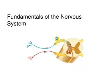
Neurons are composed of • Cell body • Processes Cell body: (perikaryon or soma) - Most neuron cell bodies located within CNS - Clusters of cell bodies in CNS are called nuclei • Few/clusters of cell bodies in PNS are called ganglia • Rough ER called Nissl bodies.
Processes - Cellular processes are called either tracts(in CNS) or nerves(in PNS) - Dendrites - have large surface area to receive chemical signals as well as conduct electrical signals (graded potentials) • Axons - single in each neuron, transmit graded potential away from cell body to axonal terminal (generates action potential) • Axon hillock arises from the cell body into the axon (graded potential) • Profuse branching at end of axon called terminal branches or telodendria. At the tips there are bulbous structures storing neurotransmitters.
Substances travel continuously up and down the axon. Anterograde: substances move from cell body to terminal axon Retrograde: substances move from terminal axon to cell body. Plasma membrane of axon is axolemma.
- Electrochemical signals transmitted with the aid by myelin sheath (protein-lipoid) which insulates nerve fibers (long axons) and increases the transmission - Nodes of Ranvier also aid in the transmission of nerve impulses. - saltitory conduction - Myelinated processes form the white matterof nervous tissue and unmyelinated processes form the gray matterof nervous tissue.
In the spinal cord, gray matter forms the H- shape in the center while white matter surrounds it. • In the brain, gray matter forms the thin outer cortex with white matter filling the inside
Classification of neurons: Structural Functional Structural: Neurons are classified as to how many processes extend from their cell body Multipolar: three or more processes extend from cell body - posses single axon - most common type of neuron in the CNS
Bipolar: two processes (axon and dendrite) extend from opposite sides of neuron. • rare in adult but may be found in retina and olfactory mucosa (special senses). Unipolar: one process extending from cell body and forms central and peripheral processes - Central process associated with secretory region • Peripheral process associated with sensory region (receptor). • Found in ganglia of PNS where they function as sensory neurons
Functional: Sensory (afferent) neurons - transmit impulses from sensory receptors toward CNS Unipolar neurons - skin or internal organs to CNS for interpretation Bipolar neurons - special sense organs, retina
Motor (efferent) neurons- carry impulses away from CNS to organs Multipolar neurons - cell body located within CNS and neurons form neuromuscular junctions with effector (muscle or gland) cells Association neurons (interneurons) - transmit impulses within CNS (usually sensory to motor); found in CNS only; mostly multipolar and 99% of neurons in body. Also called connecting neurons.
Neurophysiology/Electrophysiology: Resting Membrane Potential (RMP) Positive charge on the outside Negative charge on the inside These electrical charges are in the form of potential energy which is measured in millivolts (-70mV in the plasma membrane) The (-) in -70 mV refers to the inside of the membrane being more negative than the outside This membrane is said to be polarized
The energy in the resting membrane is likened to energy stored in a battery. Flow of electrical charge is called current. In the membrane it flows by way of ions instead of electrons (electricity) In a resting membrane (-70 mV) three Na+ ions are pumped out for every two K+ pumped in. This creates a more negative situation on the inside: Sodium/Potassium Pump
Ion channels: Leakage Gated Leakage channels are always open. Gated channels open and close in response to a stimulus. Stimulus: Voltage Chemical Mechanical pressure Light
Voltage gated ion channels will respond to the change in membrane potential (voltage). Excitability. • Chemically gated ion channels respond to the presence of a specific chemical stimulus such as hormone, neurotransmitter, Ca+ • The stimulus will result in a graded potential that will either cause the neuron to fire (depolarization) or not (hyperpolarization). Impulse traveling over a long distance (axon) is called an action potential
Depolarization: Reduction in membrane potential. When the membranes becomes less negative on the inside (moves closer to 0). Increases the probability the nerve will fire. i.e.: -70 to -65 mV -70 to +30 mV Hyperpolarization: Increase in membrane potential. When the membrane becomes more negative on the inside. Decreases the probability the nerve will fire. i.e.: -70 to -90 mV.
Depolarization: Voltage gated Na+ channels open and Na+ rushes in. -70 mV to 0mV to +30 mV. Action Potential initiated. (all or none)
Repolarization: Na+ channels close, K+ channels open and K+ is pumped out. Restores electrical conditions not original ionic distribution of resting state. It is the activation of the sodium-potassium pump that reestablishes ionic distribution +30mV to -70m. Synapse: Junction between nerve cells and effector cells. Axodendritic:
Presynaptic neuron: impulse firing away from cell body towards the synapse (sender) Postsynaptic neuron: impulse away from synapse. (receiver) Chemical synapses uses a substance called a neurotransmitter to get information across the synaptic cleft.
1- Calcium channels open in the presynaptic axonal terminal. Impulse reached axonal terminal causing a rush of Ca+ in from the extracellular fluid 2- Ca+ causes the synaptic vesicles filled with neurotransmitter to migrate and fuse with axonal membrane (exocytosis) 3- Ca+ is pumped out of terminal end 4- Neurotransmitter binds to postsynaptic receptors. 5- Ion channels open in postsynaptic membrane causing changes in membrane potential (action potential ?)
Classes of Neurotransmitters: • Acetylcholine: excitatory (skeletal muscle) • Biogenic amines • Dopamine, Norepinephrine, and epinephrine (feel good catecholamines) • Serotonin: mood, sleep, appetite & anger (inhibitory) • Histamine • Amino acids • GABA (gamma-aminobutyric acid) (inhibitory) • Glutamate: excitatory • Peptides • Endorphins and enkephalins: inhibitory (opioids)