Download
1 / 113
1.14k likes | 1.18k Views
Genetic Diseases. Dr. Joseph de Nanassy Associate Professor, PALM, uOttawa Chief of Anatomical Pathology, CHEO Site Chief of Laboratory Medicine, CHEO jdenanassy@cheo.on.ca 613-737-7600 x 2897. Objectives. Develop a basic understanding of the genetic apparatus

E N D
GeneticDiseases Dr. Joseph de Nanassy Associate Professor, PALM, uOttawa Chief of Anatomical Pathology, CHEO Site Chief of Laboratory Medicine, CHEO jdenanassy@cheo.on.ca 613-737-7600 x 2897
Objectives • Develop a basic understanding of the genetic apparatus • Comprehend definitions of major genetic abnormalities • Correlate molecular abnormalities and genetic defects
Outline I. Definitions Genetic code Chromosomes, Genes, Cell Division Molecular mechanisms II. Abnormal fetal development Malformations, deformations, dysplasias, disruptions III. Perinatal pathology Birth defects Metabolic disorders
Nucleus • DNA: arranged in chromosomes (network of granules = nuclear chromatin) • RNA: spherical intranuclear structure(s) - nucleolus / nucleoli
Genetic Code • A series of messages contained in the chromosomes • This code regulates cell functions by way of directing the synthesis of cell proteins • The code corresponds to the structure of the DNA • The code is transmitted to new cells during cell division
Chromosomes ☺ Exist in pairs – homologous: 22a + 1s ☺ Composed of double coils of DNA ☺ Basic unit: nucleotide phosphate group deoxyribose sugar base: purine (A, G) pyrimidine (T, C)
Genes ☺ A locatable region of genomic sequence, corresponding to a unit of inheritance ☺ A union of genomic sequences encoding a coherent set of potentially overlapping functional products; i.e. genes are one long continuum (2007) ☺ Determine cell properties, both structure and functions unique to the cell
Genome ☺ Sum total of all genes contained in a cell’s chromosomes ☺ Identical in all cells ☺ Not all genes are expressed in all cells ☺ Not all genes are active all the time ☺ May code for enzymes or other functional proteins, structural proteins, regulators of other genes
Gene Product ☺ A protein or RNA specified by a gene ☺ Transcribed into mRNA in the nucleus ☺ Translated through tRNA and cytoplasmic ribosomes into protein
Human Genome ☺ 3 billion+ pairs of DNA nucleotides ☺ ~ 50,000 – 100,000 genes ☺ Protein-coding Genes = <10% (2%) of human genome ☺ Exons: parts of the DNA chain that code for specific proteins ☺ Introns: the parts in-between the exons ☺ Both exons and introns are transcribed but only the exons are translated (introns are removed from mRNA before leaving nucleus) ☺”Junk DNA”: no obvious function but 80% expressed
Sex chromosomes ☺ Genetic sex = composition of X and Y ☺ Large X: many genes, many activities ☺ Small Y: almost entirely male sexual diff. ☺ Female: XX, male XY ☺ One X randomly inactivated and nonfunctional after first week of embryonic development ☺ Same inactivated X in descendant cells
Y chromosome ☺ Stains with some fluorescent dyes - bright fluorescent spot in the nucleus ☺ Normal female: sex chromatin body but no fluorescent spot ☺ Normal male: fluorescent spot but no sex chromatin body
Cell Division ☺ Mitosis: somatic cells (PMAT) Daughter cells have the same number of chromosomes as the parent cell. ☺ Meiosis: gametogenesis (1st and 2nd div) Number of chromosomes reduced by half.
Chromatids ☺ Before mitosis, the DNA chains duplicate to form new chromosome material. The duplicated chromosome material lies side by side = two sister chromatids. Mitosis = the process by which conjoined chromatids separate into sister chromatids and move into new daughter cells.
Mitosis ☺ Interphase: DNA duplication to form chromatids just before mitosis ☺ Prophase: centriole migration, mitotic spindle ☺ Metaphase: chromosomes line up in centre, chromatids still joined at centromere ☺ Anaphase: chromosomes separate into sister chromatids ☺ Telophase: sister chromatids form new chromosomes, new nuclear membranes form, cytoplasm divides
Meiosis ☺ First meiotic division interphase: duplication of chromosomes to form paired chromatids ☺ Prophase 1 of meiosis: homologous chromosomes lie side by side over entire length = synapse. Interchange of segments of homologous chromosomes = crossover. 2 Xs side by side just like the autosomes. X and Y end-to-end: no crossover.
Meiosis ☺ Metaphase 1: paired homologous chromosomes align at the equatorial plate ☺ Anaphase 1: homologous chromosome pairs migrate to opposite poles of the cell; each chromosome is composed of two chromatids, the chromatids are not separated ☺ Telophase 1: two new daughter cells form; each contains half the chromosome number = reduction of chromosomes by half; interchange of genetic material occurred during synapse
Meiosis ☺ Second meiotic division = mitotic division Prophase 2: DNA does not replicate Metaphase 2: chromosomes align at the equatorial plate Anaphase 2: sister chromatids migrate separately Telophase 2: four haploid cells (half the normal number of chromosomes)
Gametogenesis ☺ Gonads: testes, ovaries; contain ☺ Precursor cells or germ cells; mature into ☺ Gametes: sperm, ova; in gametogenesis ☺ Spermatogenesis, oogenesis
Oogenesis vs. spermatogenesis ☺ One ovum (+ 3 polar bodies) vs. four spermatozoa ☺ Oocytes formed before birth vs. continuous spermatogenesis (‘fresh’ sperm) Prolonged Prophase 1 until ovulation – more frequent congenital abnormalities in ova of older women (longer exposure to potentially harmful environmental influences until meiotic division resumes at ovulation)
Genes and Inheritance ☺ Locus: specific site of a gene on the chromosome. Since the chromosomes exist in pairs, genes are also paired. ☺ Alleles: alternate forms of a gene can occupy the same locus (homozygous, heterozygous) ☺ Recessive gene: expressed only when homozygous ☺ Dominant gene: expressed whether homozygous or heterozygous, both expressed when co-dominant ☺ Sex-linked gene: only X-linked in males, most are recessive, hemizygous (no allele on Y)
Gene Imprinting ☺ Genes occur in pairs on homologous chromosomes, one from each parent ☺ Different effects of gene whether ♀ or ♂ ☺ Genes modified during gametogenesis ☺ Gene imprinting: additional methyl groups added to DNA molecules ☺ Basic structure unchanged; in some diseases different expression (behaviour) depending on parent of origin: hereditary disease as a result of imprinting
Genetic Engineering ☺ Insertion of a gene encoding a desired product (e.g. insulin) into a bacterium ☺ Bacterial gene spliced enzymatically, recombinant DNA inserted into plasmid (circular DNA segment in bacterium), dividing bacterial population produces desired protein
Gene Therapy ☺ Normal gene inserted into defective cell ☺ Compensates for the missing or dysfunctional gene, in somatic cells only ☺ Can be inserted into mature cell (ly) ☺ Can be inserted into stem cell (bone marrow) ☺ Used to treat e.g. ADA deficiency, CF, …
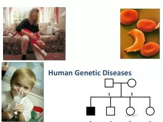
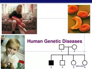
Congenital / Hereditary Diseases ☺ Congenital: present at birth ☺ Hereditary (genetic): result of chromosome abnormality or defective gene
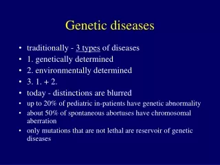
Causes of malformations • Chromosomal abnormalities • Gene abnormalities • Intrauterine injury (e.g. drugs, radiation, infection, environmental, etc) • Environmental effect on genetically predisposed embryo
Chromosomal abnormalities ☺ Nondisjunction: failure of homologous chromosomes in germ cells to separate from one another during 1st or 2nd meiotic division ☺ Sex chromosomes or autosomes ☺ Extra chromosome: trisomy (24 or 47) Absent chromosome: monosomy (22 or 45)
☺ Chromosome Deletion: Broken piece of chromosome is lost from cell ☺ Translocation: Not lost, just misplaced and attached to another chromosome - reciprocal: between two nonhomologous chromosomes (no loss or gain of genetic material - no loss of cell function) - in germ cells: deficient or excess chromosome material – abnormal zygote
Autosomal abnormalities ☺ Loss of genetic material: aborted embryo ☺ Deletion of gene: congenital anomalies ☺ Trisomy: syndromic, e.g. 21, 18, 13
T21 causes • Nondisjunction during gametogenesis (95%) • Translocation (few) • Nondisjunction in zygote (rare)