Download
1 / 34
340 likes | 577 Views
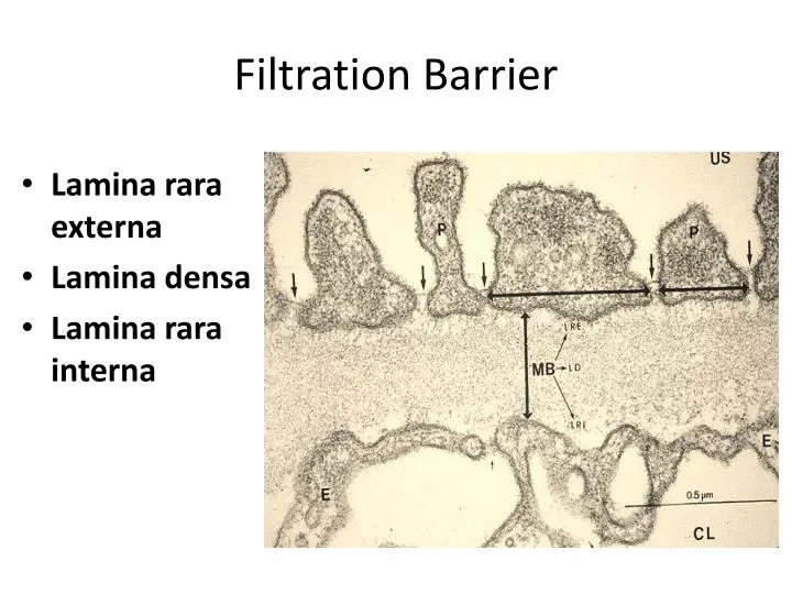
Filtration Barrier. Lamina rara externa Lamina densa Lamina rara interna. Glomerular Filtration. Filtering of blood in the renal corpuscle Glomerular filtrate – fluid that seeps from the glomerular capillaries into the Bowman’s space. Renal Tubule. Proximal convoluted tubule

E N D
Filtration Barrier • Lamina rara externa • Lamina densa • Lamina rara interna
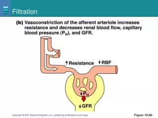
Glomerular Filtration • Filtering of blood in the renal corpuscle • Glomerular filtrate – fluid that seeps from the glomerular capillaries into the Bowman’s space
Renal Tubule • Proximal convoluted tubule • Loop of Henle • Thick descending limb • Thin descending and ascending limbs • Thick ascending limb • Distal convoluted tubule • Collecting tubules and ducts
Types of Nephrons • Short-looped (cortical) - majority • Renal corpuscles located in outer portions of cortex • Short Henle’s loop (no thin ascending limb) • Long-looped (juxtamedullary) • Renal corpuscles are near the corticomedullary junction • Long Henle’s loop (go deep into medulla, have thin ascending limb)
Juxtaglomerular Apparatus • Microscopic structure in the kidney which regulates the function of each nephron • Found between the vascular pole of the renal corpuscle and the returning DCT of the same nephron • Function: regulates filtrate formation and systemic BP
Juxtaglomerular Apparatus • 3 cells: • Juxtaglomerular cells • Mesangial cells • Macula densa cells - area of closely packed specialized columnar cells lining the wall of the distal tubule • Sense changes in solute concentration and flow rate of filtrate
BLADDER AND URINARY PASSAGES • Renal calyces • Renal pelvis • Ureter • Urinary bladder • Same histologic structure • Walls of ureter becoming gradually thicker as proximity to the bladder increases
BLADDER AND URINARY PASSAGES • Function: • Store the urine formed in the kidneys and conduct it to the exterior
BLADDER AND URINARY PASSAGES • Mucosa: • Transitional epithelium • Lamina propria: loose to dense CT • Muscular layer – OCIL • Adventitia or serosa • Only upper part of bladder covered by peritoneum
URETHRA • Terminal portion of the urinary system • Function: • Passageway for urine • Males: passageway for urine and sperms
Male Urethra • 20 cm in length • 3 segments: • Prostatic urethra • Membranous urethra • Spongy urethra (penile, cavernous) • Terminates in the external urethral orifice (meatus)
Female Urethra • Shorter (4 cm) • Closely attached to anterior wall of vagina • Opens directly in front of the vaginal opening on the vestibule of the external genitalia
Female Urethra • Mucosa • Transitional → stratified squamous non-keratinized • Lamina propria: loose CT, mucus-secreting urethral glands • Muscular layer: circularly arranged smooth muscle cells
Female Urethra • Distal segment is surrounded by circularly-arranged striated (voluntary) muscle fibers (sphincter urethrae muscle – external sphincter)