Download
1 / 22
220 likes | 497 Views
Filtration. Figure 19-8b. Filtration. Figure 19-8c. GFR Regulation. Myogenic response Similar to autoregulation in other systemic arterioles

E N D
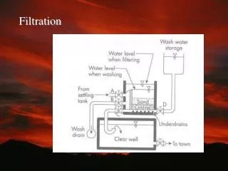
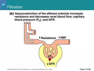
Filtration Figure 19-8b
Filtration Figure 19-8c
GFR Regulation • Myogenic response • Similar to autoregulation in other systemic arterioles • When smooth muscle in wall of arteriole stretches, stretch-sensitive ion channels open, and the muscle cells contract. Vasoconstiction increases resistance and slows flow. This keeps GFR constant. • Tubuloglomerular feedback • Hormones and autonomic neurons • By changing resistance in arterioles (sympathetic stimulation causes vasoconstriction) • By altering the filtration coefficient
Juxtaglomerular Apparatus Figure 19-9
Tubuloglomerular Feedback Distal tubule Efferent arteriole Glomerulus Bowman’s capsule GFR increases. 1 Proximal tubule Macula densa 2 Flow through tubule increases. 4 1 5 3 Flow past macula densa increases. Afferent arteriole Granular cells 3 2 2 4 Paracrine diffuses from macula densa to afferent arteriole. 5 Afferent arteriole constricts. Resistance in afferent arteriole increases. Collecting duct Hydrostatic pressure in glomerulus decreases. Loop of Henle GFR decreases. PLAY Animation: Urinary System: Glomerular Filtration Figure 19-10
Tubuloglomerular Feedback Distal tubule Efferent arteriole Glomerulus Bowman’s capsule GFR increases. 1 Proximal tubule Macula densa 2 Flow through tubule increases. 4 1 5 3 Flow past macula densa increases. Afferent arteriole Granular cells 3 2 2 4 Paracrine diffuses from macula densa to afferent arteriole. 5 Afferent arteriole constricts. Resistance in afferent arteriole increases. Collecting duct Loop of Henle Figure 19-10, steps 1–5 (2 of 4)
Reabsorption • Transepithelial transport • Substances cross both apical and basolateral membrane • Paracellular pathway • Substances pass through the junction between two adjacent cells
Reabsorption Sodium reabsorption in the proximal tubule Figure 19-12
Reabsorption Sodium-linked glucose reabsorption in the proximal tubule Figure 19-13
Reabsorption • Urea • Passive reabsorption • Plasma proteins • Transcytosis PLAY Animation: Urinary System: Early Filtrate Processing
Reabsorption Saturation of mediated transport Figure 19-14
Reabsorption Glucose handling by the nephron Figure 19-15a
Reabsorption Figure 19-15b
Reabsorption Figure 19-15c
Secretion • Transfer of molecules from extracellular fluid into lumen of the nephron • Active process • Secretion of K+ and H+ is important in homeostatic regulation • Enables the nephron to enhance excretion of a substance
Excretion • Excretion = filtration – reabsorption + secretion • Clearance • Rate at which a solute disappears from the body by excretion or by metabolism • Non-invasive way to measure GFR • Inulin and creatinine used to measure GFR
Excretion The relationship between clearance and excretion Figure 19-17a
Excretion Figure 19-17b
Excretion Figure 19-17c
Micturition The storage of urine and the micturition reflex Figure 19-18a
Micturition 1 2 3 Stretch receptors fire. Parasympathetic neurons fire. Motor neurons stop firing. Smooth muscle contracts. Internal sphincter passively pulled open. External sphincter relaxes. (b) Micturition Higher CNS input may facilitate or inhibit reflex. Stretch receptors Sensory neuron 1 Parasympathetic neuron 2 3 + – Motor neuron Tonic discharge inhibited Internal sphincter 2 3 External sphincter Figure 19-18b