Download

1 / 26
340 likes | 861 Views
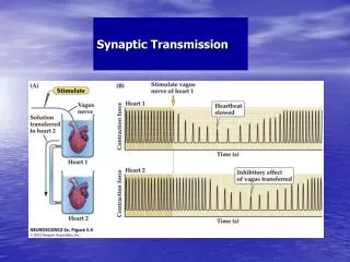
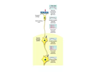
Synaptic Transmission. 14.5.12. All or None Action Potential. AP in neurons is unidirectional.

E N D
Synaptic Transmission 14.5.12
AP in neurons is unidirectional • After generating an axon potential at trigger zone, it begins to propagate to neighboring segments of the membrane and depolarize them to threshold triggering action potentials in the next neighboring area and so on • This propagation is unidirectional from axon hillock to axon terminal because in the case of the neuron, the proximal segment just traversed by the action potential just enters a refractory period and thus becomes inexcitable
Absolute Refractory Period • The period when a recently activated patch of a membrane is completely refractory (meaning “stubborn” or unresponsive) to further stimulus is known as “absolute refractory period” • Once the voltage gated Na+ channels are triggered to open at threshold, they cannot open again in response to another stimulus no matter how strong until they pass through their “closed and not capable of opening” conformation and they are reset to their “closed and capable of opening” conformation • Absolute refractory period lasts the entire time from threshold to depolarization peak and until return to resting potential
Relative Refractory Period • Following the absolute refractory period is a “relative refractory period”, during which a second action potential can be produced only by a triggering event considerably stronger than usual • The voltage gated K+ channels that opened at the peak of action potential are slow to close. During this time, the resultant less than normal Na+ entry in response to another triggering event is opposed by K+ still leaving through its slow to close channels during hyperpolarization • Thus a greater depolarizing triggering event than normal is needed to offset the persistent hyperpolarizing outward movement of K+ and bring the membrane to threshold during the relative refractory period
2 ways to increase AP propagation speed • Increase internal diameter of axon which decreases the internal resistance to ion flow • Increase the resistance of the plasma membrane to charge flow by insulating it with myelin.
Speed of Nerve Impulses • Impulses travel very rapidly along neurons. The presence of a myelin sheath greatly increases the velocity at which impulses are conducted along the axon of a neuron. In unmyelinated fibres, the entire axon membrane is exposed and impulse conduction is slower.
Saltatory Conduction Saltatory Conduction: Action potentials jump from one node to the next as they propagate along a myelinated axon.
Myelin acts as an insulating sheath allowing an action potential to spread along the axon until it gets to a node of Ranvier which is a bare portion of axon without mylin. As a result action potential jumps from one node to the next and so on. This is called saltatory conduction
Properties of Graded vs Action Potential • Graded Potential • Amplitude varies with size of initiating event • Can be summed • Has no threshold • Has no refractory period • Is conducted decrementally i.e. amplitude decreases with distance • Can be a depolarization or a hyperpolarization • It begins with a stimulus (chemical, electrical, mechanical) or a synapse • Mechanism depends on ligand gated channels, or other physical or chemical chages • Action Potential • All or None. Once the membrane is depolarized to threshold, amplitude is independent of size of initiating event • Cannot be summed • Has a threshold which is usually 15mV depolarized relative to resting potential • Has a refractory period • Is conducted without decrement • Is a depolarization initially than a hyperpolarization • Initiated only by a graded potential • Mechanism depends on voltage gated channels
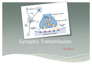
Synaptic Transmission • Synapse is the place where information is transferred from one neuron to other • Two types Electrical synaptic transmission Chemical synaptic Transmission
Electrical Synaptic Transmission • These synapses refer to gap junction between two nerve cells • These synapses separate two adjoining neurons by a few nanometers • The cytoplasm of one cell is connected to the next cell through channels named connexons • Current can flow through these channels either way, thus depolarization and hyperpolarization can spread from one cell to to next cell instantaneously • Gap junction are good in spreading electrical signals through networks of interconnected neurons and are effective in developing synchronic activity in clusters of neurons
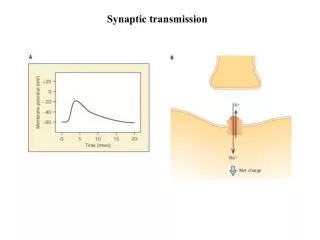
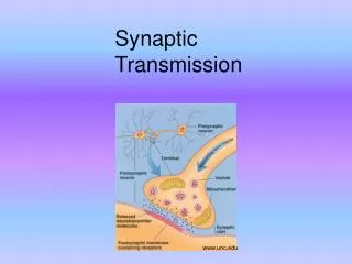
Chemical Synaptic Transmission • Here there is no continuity of cytoplasm at all and the direction of signal transmission is unidirectional • The gap between two cells is extracellular space and named the “synaptic cleft” • When the action potential reaches axon terminal, intracellular Ca2+ increases by opening of voltage gated calcium channels • The increased levels of Ca++ triggers fusion of synaptic vesicles wherein neurotranmitters are stored within neuronal membrane • The released neurotransmitters bind to specific receptors of the post synaptic cell membrane and change the membrane potential
Functional anatomy of the synapses The synapse is the point of communication between two neurons that operate sequentially.
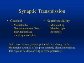
Activation of the Postsynaptic cell: An excitatory postsynaptic potential (EPSP) is a graded depolarization that moves the membrane potential closer to the threshold for firing an action potential (excitement). An inhibitory postsynaptic potential (IPSP) is a graded hyperpolarization that moves the membrane potential further from the threshold for firing an action potential (inhibition).
Interaction of Excitatory and Inhibitory synapses Panel 1: Two distinct, non-overlapping, graded depolarizations. Panel 2: Two overlapping graded depolarizations demonstrate temporal summation. Panel 3: Distinct actions of stimulating neurons A and B demonstrate spatial summation. Panel 4: A and B are stimulated enough to cause a suprathreshold graded depolarization, so an action potential results. Panel 5: Neuron C causes a graded hyperpolarization; A and C effects add, cancel each other out.
Modifications of synaptic transmission by drugs and disease Possible drug effects on synaptic effectiveness: A. release and degradation of the neurotransmitter inside the axon terminal. B. increased neurotransmitter release into the synapse. C. prevention of neurotransmitter release into the synapse. D. inhibition of synthesis of the neurotransmitter. E. reduced reuptake of the neurotransmitter from the synapse.
Excitable Muscle Cells: Motor unit • When a single nerve enters a muscle it splits and makes neuromuscular junctions (NMJs) with several muscle fibers • A nerve and the muscle cells (fibers) it makes NMJs with is called a motor unit • When the nerve fires the whole motor unit is stimulated and the muscle cells contract together
Excitable Muscle Cells • Single stimuli usually release enough acetylcholine in the NMJs of the motor unit to produce action potentials in the muscle membranes • This will cause the muscle to contract after a short delay • Order of events: ACh release -> muscle action potential -> Ca release -> contraction
Tension developed by Muscle Fiber • Tension developed by a single muscle fiber depends on • Length of the fiber at the onset of contraction • Frequency of Stimulation
Twitch contraction • With a single stimulus (one action potential in the nerve that excites a muscle) after an activation delay of 5msec, the muscle fiber will contract. • The tension in the muscle fiber rises rapidly to a maximum and then return to its resting tension This is called a twitch contraction or a twitch
A second stimulus applied before the muscle fiber has completely relaxed, induces another contraction that adds to the first, the sum of the tensions being greater than that of a single twitch • Maximum tension that can be developed in a muscle fiber due to high frequency of stimulation is called “ tetanic contraction”