Download

1 / 43
440 likes | 659 Views
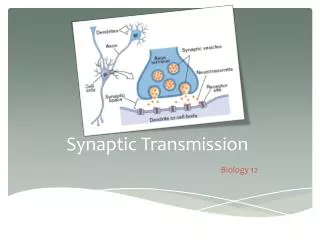
Synaptic transmission 1. Synaptic Transmission. Expiratory neuron (top trace) and inspiratory neuron (bottom trace) were labeled with dye during intracellular recording from the ventrolateral medulla. Clearly, activity in each one of these cells affects activity in the other one. Outline.

E N D
Synaptic Transmission • Expiratory neuron (top trace) and inspiratory neuron (bottom trace) were labeled with dye during intracellular recording from the ventrolateral medulla. Clearly, activity in each one of these cells affects activity in the other one.
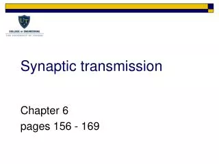
Outline • A. Electrical synapses • B. Overview of chemical synapses • C. Synaptic transmission via acetylcholine • D. Diversity of chemical synapses • E. Norepinephrine/serotonin and depression
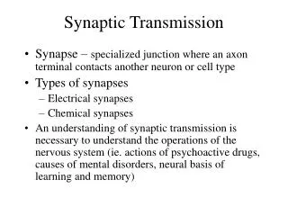
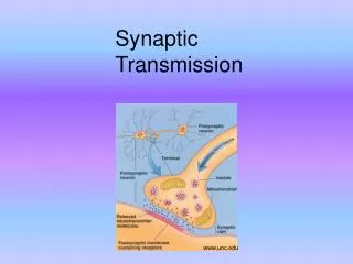
Synapses • Cellular junctions where signals are transmitted from neurons to target cells • These are communicating junctions • Target cells: Other neurons, muscle cell, gland cells • Two types of communicating junctions or synapses: Electrical synapses via gap junctions, chemical synapses involving neurotransmitters
Electrical synapse and gap junctions • Recall that this involves channels comprised of connexons that link cells
Gap junctions • A patch where cells are separated by a narrow gap of 2-4 nm • Connexons, Connexins • Each connexon is comprised of six identical subunits (connexins) • Permeability of junction mediated by conformation of the connexons
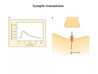
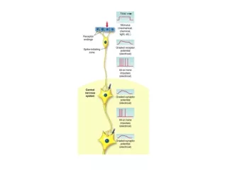
Impulse transmission across synapses • Some terminology: • Presynaptic cell • Neuron carrying action potential • Postsynaptic cell • Target cell receiving signal Transmission of signal can result in a depolarization of the postsynaptic cell - an excitatory postsynaptic potential (EPSP), Or hyperpolarization, or simply stabilization, of the membrane potential of the postsynaptic cell – an inhibitory postsynaptic potential (IPSP)
Impulse transmission across electrical synapses is almost instantaneous • Ions move directly from presynaptic cell to postsynaptic cell via gap junctions • Transmission occurs in a few microseconds • Over a hundred times faster than in chemical synapses
Transmission of an action potential across an electrical synapse
Under what circumstances are electrical synapses important? • Invertebrate escape responses • Also escape responses in vertebrates such as goldfish • Large number of electrical synapses in fishes living at low temperature • Can also be used to electrically couple groups of cells so they are synchronized
Summary • Transmission of signals across electrical synapses is rapid • This involves movement of ions via gap junctions • Used when rapid conduction of signals is essential or to synchronize cells
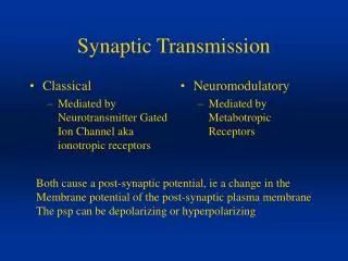
Chemical synapses • Overall: • Action potential of presynaptic cell causes release of neurotransmitter into the synaptic cleft • Binding of neurotransmitter to postsynaptic cell results in a depolarization at excitatory synapses (an excitatory postsynaptic potential EPSP) or stabilization or hyperpolarization at inhibitory synapses (an IPSP).
Step 2 N Ca++ channels
How vesicle fusion occurs: Reserve pool of vesicles is free in synaptic terminal – but these have to undergo docking and priming to be ready to release Some vesicles are attached to the presynaptic membrane by connections between specific proteins on vesicle and counterparts on presynaptic membrane- at least 6 different proteins are believed to be involved. These are primed. They have joined the readyreserve pool. To enter the ready-to-release pool, a primed vesicle must be docked by becoming associated with n-type Ca++ channels at the presynaptic membrane. Depolarization opens the Ca++ channels – tiny geysers of Ca++ occurs at that vesicle’s location – this Ca++ causes vesicle fusion – transmitter is released into synaptic cleft.
Some of the “players” in (a) docking (b) fusion preparation and (c) Ca++-sensitive exocytosis
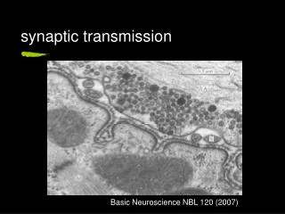
Freeze-Fracture view of vesicle release Docking proteins and N-type Ca++ channels are visible in the picture at left. In the picture at right we are looking into the mouths of several open vesicles.
Vesicle Membrane Conservation: a kiss-and-run process - the motor protein dynamin pinches and the coating protein clathrin forms a cage around the membrane…
Toxins and synaptic vesicle fusion… • Synaptobrevin and SNAP-25 are targets of the clostridial neurotoxins: tetanus toxin acts in the Central Nervous System (CNS) and botulinum toxin acts at neuromuscular synapses – paralysis is caused by blockage of transmitter release. • Neurexin is targeted by a-latrotoxin, the black widow spider toxin, which induces massive transmitter release independent of Ca++ levels.
Transmission of an action potential across chemical synapse Most of the synaptic delay (1-2 msec) is due to the time it takes to organize the presynaptic processes
Part C: Transmission via acetylcholine A fairly well-understood example
I. Storage of acetylcholine (ACh) in synaptic vesicles • 40 nm diameter membrane bounded vesicles • Contain 1000 to 10,000 molecules of acetylcholine • A single axon terminus may contain a million or more vesicles contacting the target cell at several hundred points
What neuromuscular synapse anatomy reveals: • The area of contact at the neuromuscular synapse is very extensive. • Glia cover the area of the synapse. • Highly specialized regions exist in both cells: • The neurons have the large accumulations of synaptic vesicles and associated release system • The muscle cell has an accumulation of receptors and other response elements that will allow the signal to spread over the membrane and within the cell.
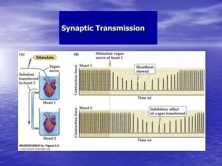
Acetylcholine (ACh) and the neuromuscular synapse: • In 1921 Otto Loewi showed that ACh was released at synapses (and also into the saline) by the vagus nerve: andtransfer of the solution slowed the heartbeat of a second frog heart.
Acetylcholine ACh is a transmitter that is in a class by itself: • It is synthesized in terminals from acetyl CoA and choline by choline acetyltransferase. • It is packaged in vesicles in the axon terminals. • It can bind to two distinct receptor types: nicotinic and muscarinic. Nicotinic receptors are seen in the skeletal muscle synapse and at synapses within the CNS. Muscarinic receptors for ACh are also seen in the CNS and at parasympathetic synapses on target tissues. • After release, ACh is degraded by the enzyme acetylcholinesterase into acetate and choline. • The choline is taken back into the terminal by Na+-driven facilitated uptake.
Synthesis of acetylcholine • Takes place in cytosol of axon terminals
Accumulation of acetylcholine in synaptic vesicles • Involves active transport Vacuolar-type H+ATPase
Accumulation of acetylcholine • V type ATPase in vesicle membrane is used to reduce vesicle pH • Low vesicle pH powers a proton/neurotransmitter (NT) antiporter