Download
1 / 52
520 likes | 670 Views
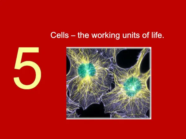
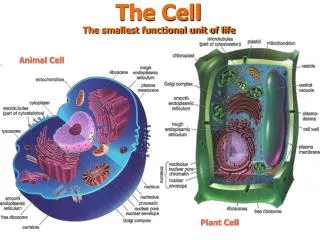
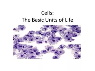
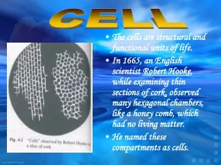
Introduction to the cell 1. Cells are the smallest units of life 2. Cells divide to produce new cells 3. Cells are microscopic 4. Eukaryotic cells contain organelles 5. Cells are diverse in size, shape, function 6. Cells arose from a common ancestor

E N D
Introduction to the cell 1. Cells are the smallest units of life 2. Cells divide to produce new cells 3. Cells are microscopic 4. Eukaryotic cells contain organelles 5. Cells are diverse in size, shape, function 6. Cells arose from a common ancestor Post-genomic cell biology exploits organisms whose genomes are sequenced
Microscopes Resolving Power - Resolution R ~ 1/2 l R = 0.6 l/NA
Breakthroughs 2002: Frozen Images Thirty years ago, researchers pitched the idea of reconstructing a three-dimensional picture from electron micrographs. Today, cryoelectron tomography (cryo-ET) has overcome a series of technical obstacles to emerge as a breakthrough technique for viewing structures inside intact cells. Actin in the act. Cryoelectron tomography captures new views of cellular components, such as these actin filaments. CREDIT: O. MEDALIA ET AL.SCIENCE298, 1209 (2002)26 December 2002 Biologists have long been able to capture the molecular structure of single proteins in cells, using techniques such as x-ray crystallography. But they haven't had a good way to get a 3D look at midsize organelles (~5 nm), such as the protein-packaging Golgi apparatus or energy-producing mitochondria, especially without removing them from their native environment. Cryo-ET fills this resolution gap and gives scientists a way to link atomic-level detail to whole cell organization. Cryo-ET works something like a doctor's computerized tomography scan. Penetrating beams of electrons create two-dimensional image slices that a computer assembles into a 3D image. Cells are flash-frozen and do not need to be fixed or their membranes disrupted. For years, the problem with cryo-ET has been that too much radiation causes structures to degrade. Long, steady progress has solved many of the early snags. Autorotation of the specimen through a range of imaging angles and better calibration of the microscope stage have dramatically reduced exposure time. Improved clarity by reduced scattering of the electrons allows the viewing of thicker specimens. Advances in cryosectioning, slicing up the specimen in layers, have also enabled this technique to be used with thicker samples. This year, cell imagers used cryo-ET to catch the first glimpse of actin filaments in the act, braced against the edge of the cell membrane. They also captured the first view of spatial arrangement of tubules and receptors in the sarcoplasmic reticulum, the components responsible for the chemical cascade that sets off a muscle contraction. And efforts are currently under way to create the first detailed 3D map of the spatial relationship of all the organelles in a eukaryotic cell.
Breakthroughs 2002: Frozen Images Thirty years ago, researchers pitched the idea of reconstructing a three-dimensional picture from electron micrographs. Today, cryoelectron tomography (cryo-ET) has overcome a series of technical obstacles to emerge as a breakthrough technique for viewing structures inside intact cells. Actin in the act. Cryoelectron tomography captures new views of cellular components, such as these actin filaments. CREDIT: O. MEDALIA ET AL.SCIENCE298, 1209 (2002)26 December 2002 Biologists have long been able to capture the molecular structure of single proteins in cells, using techniques such as x-ray crystallography. But they haven't had a good way to get a 3D look at midsize organelles (~5 nm), such as the protein-packaging Golgi apparatus or energy-producing mitochondria, especially without removing them from their native environment. Cryo-ET fills this resolution gap and gives scientists a way to link atomic-level detail to whole cell organization. Cryo-ET works something like a doctor's computerized tomography scan. Penetrating beams of electrons create two-dimensional image slices that a computer assembles into a 3D image. Cells are flash-frozen and do not need to be fixed or their membranes disrupted. For years, the problem with cryo-ET has been that too much radiation causes structures to degrade. Long, steady progress has solved many of the early snags. Autorotation of the specimen through a range of imaging angles and better calibration of the microscope stage have dramatically reduced exposure time. Improved clarity by reduced scattering of the electrons allows the viewing of thicker specimens. Advances in cryosectioning, slicing up the specimen in layers, have also enabled this technique to be used with thicker samples. This year, cell imagers used cryo-ET to catch the first glimpse of actin filaments in the act, braced against the edge of the cell membrane. They also captured the first view of spatial arrangement of tubules and receptors in the sarcoplasmic reticulum, the components responsible for the chemical cascade that sets off a muscle contraction. And efforts are currently under way to create the first detailed 3D map of the spatial relationship of all the organelles in a eukaryotic cell.
Macromolecular Architecture in Eukaryotic Cells Visualized by Cryoelectron Tomography Ohad Medalia, Igor Weber, Achilleas S. Frangakis, Daniela Nicastro, Günther Gerisch, Wolfgang Baumeister
The Human Genome Project The Post-Genomic Era Functional Genomics Functional Proteomics Bioinformatics
Practice questions at the end of chapter I 1-8, 9, 12, 16, 19