Download

1 / 65
650 likes | 699 Views
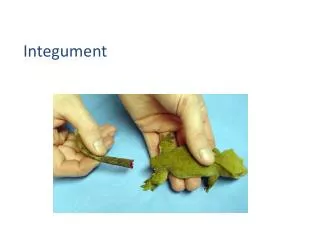
Explore the layers, functions, and cell types of the integumentary system in animals. Discover the unique features of paw pads, hair follicles, and skin-related glands. Learn about the epidermis, dermis, and hypodermis structures.

E N D
Learning Objectives • List the cell types that make up the epidermis and describe the function of each cell type. • List the five layers of the epidermis. • Describe the process of keratinization. • List the structures that constitute the dermis and describe the function of each. • List the structures of the hypodermis. • Describe the unique features of the paw pads and planum nasale. • Describe the parts of the hair follicle and explain how hair grows. • List and describe the three types of hair. • Describe the structure and location of sebaceous glands. • Differentiate between eccrine and apocrine sweat glands.
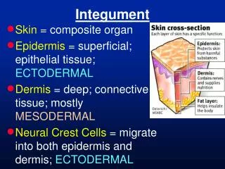
Integumentary System • Skin and related structures: • 1. Hair, • 2. hooves, • 3. paw pads • 4. horns and antlers, • 5. claws and dewclaws, • 6. noses (muzzles) • 7. skin-related glands sebaceous and sweat
Functions: • 1. prevents desiccation (drying up); • 2. reduces threat of injury; • 3. assists in maintaining normal body temperature; • 4. excretes water, salt, and organic wastes; • 5. receives and conveys sensory information; • 6. synthesizes vitamin D; stores nutrients
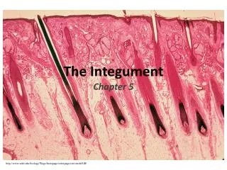
Integumentary System • Consists of three layers: • Epidermis • Dermis • Hypodermis
Epidermis Cell types: 1. Keratinocytes – produce keratin, the tough, fibrous, waterproof protein that gives skin its resiliency and strength live cells at the basement membrane, dead at the surface (stratum corneum) 2. Melanocytes– produce melanin pigment
3. Merkel cells– tactile sensory nerve endings found in the basal part of the epidermis • 4. Langerhanscells– found in stratum spinosum; • MACROPHAGE that is specific to the epidermis ( may be involved in allergic and cell-mediated immune response in skin)
Epidermal Layers • Stratum germinativum (basal layer): • Deepest layer • Consists of a single row of keratocytes attached to epithelial basement membrane • Merkel cells and melanocytes are also found in this layer
Epidermal Layers Stratum spinosum (spiny layer): Langerhans cells found in this layer Less active keratinocytes
Epidermal Layers • Stratum granulosum (granular middle layer): • Composed of two to four layers of flattened, diamond-shaped keratocytes that contain lamellated granules of glycolipids • Degeneration of nucleus and organelles…the cells die in this layer • These glycolipids play a role in helping waterproof the skin and slowing water loss across the epidermis
Epidermal Layers • Stratum lucidum (clear layer): • Found in very thick skin • Composed of a few rows of flattened dead cells
Epidermal Layers 5.Stratum corneum (horny outermost layer): • Composed of 20 to 30 rows of keratocyte “remnants” • Sometimes called horny or cornified cells
Epidermis of Hairy Skin • Hairy skin usually consists of three epidermal layers rather than five (stratum basale, stratum spinosum, and stratum corneum) • The surface of hairy skin is covered in scalelikefolds with hair in clusters of 3 follicles per scale. • Tactile Elevations: • Tactile elevation or epidermal papilla • associated with a tactile hair (touch) • (tylotrichhairs)
Epidermis 1 -5 layers • Stratum Basale • Stratum Spinosum • Stratum Granulosum • Stratum Lucidum • Stratum Corneum • Living Keratinocytes, Merkel Cells, Melanocytes • Less active keratinocytes, Langerhans cells • Dying keratinocytes • “clear layer” dead cells • Dead cells
Dermis • Composed of dense irregular connective tissue • Collagen, elastic, and reticular fibers • Also includes hair follicles, nerve endings, glands, smooth muscle, blood vessels, and lymphatics • Fibroblasts, adipocytes, and macrophages also present • Two layers: • Papillary layer • Reticular layer
Dermal Layers Papillary layer • Underneath the epithelial layer of the epidermis • Composed of loose connective tissue with loosely woven fibers and ground substance • Dermal papillae help cement the epidermis and the dermis together • Blood vessels, pain, temperature, and touch receptors also present
Dermal Layers Reticular layer • Consists of dense irregular connective tissue • Bundles of collagen fibers from papillary layer blend into those of reticular layer • Most fibrous bundles tend to run parallel to each another. • Separations between bundles represent tension lines in skin • In areas where a great deal of bending occurs, dermal folds or flexure lines are present.
Hypodermis Composed of areolar tissue containing adipose, blood and lymphatic vessels, and nerves Contains special touch receptor – the Pacinian corpuscle (sensitive to heavier pressure than Meissner's corpuscle) Fibers of hypodermis are continuous with those of dermis Hypodermal layer permits skin to move freely over underlying bone and muscle without putting tension on skin
Special Features of the Integument Pigmentation Paw Pads Planum Nasale Ergots and Chestnuts Cutaneous Pouches in Sheep
Pigmentation • Result of presence or absence of melanin granules in the extensions of melanocytes • No pigmentation if granules are concentrated around nucleus of the melanocyte • As granules move into the cellular extensions and into surrounding tissue, pigmentation becomes macroscopically apparent (you can see it) • The more granules present, the darker the pigmentation
Pigmentation Melanocyte-stimulating hormone controls dispersion of granules Keratinocytes arrange melanin on the side of the cell with greatest amount of sun exposure Acts to protect keratinocytes from exposure to damaging ultraviolet rays
Paw Pads • Thick layers of fat and connective tissue with exocrine sweat glands and lamellar corpuscles (Lamellar corpuscles, or Pacinian corpuscles, are nerve endings in the skin responsible for sensitivity to vibration and pressure.) • Outer surface is the toughest and thickest skin in the body • Often pigmented; composed of all five epidermal layers • Stratum corneum (top layer) is thicker than all other layers combined • Conical papillae can be seen covering entire pad
Planum Nasale • Top of the nose in cats, pigs, sheep, and dogs • Planum nasolabiale:the muzzle of cows and horses • Usually pigmented; aglandular except in sheep, pigs, and cows • Composed of only three epidermal layers: • Stratum germinativum, stratum spinosum, stratum corneum • Not present: stratum lucidum, stratum granulosum
Nasal Planum is composed of polygonal plaques separated by epidermal grooves.
Ergots and Chestnuts • Dark horny structures found on the legs of horses, ponies, and other members of the equine family • Thought to be vestiges of carpal and tarsal pads of second and fourth digits ("splint bones")
Cutaneous Pouches in Sheep Infoldings of skin Infraorbital, interdigital, and inguinal pouches Contain sebaceous “oil” glands Secrete a fatty yellow substance which covers and sticks to the skin when dry
Related Structures of the Integument • Hair • Hair strands and follicles • Types of hair • Glands of the skin • Sebaceous and sweat glands • Tail glands • Anal sacs • Claws and dewclaws • Hoof • Horns
Hair • Function: • maintaining body temperature by trapping layers of air (for insulation) • Dark hair, absorbs light and can contribute to warmth • camouflage
Parts: Hair shaft: visible above the skin Hair root: buried within the skin Hair follicle: anchors the hair extends from skin surface to the dermis (occasionally the hypodermis) • .
Deepest part of hair follicle expands to form a hair bulb • At the base of the hair bulb is a mound of dermal cells called thepapilla.
Hair Strands Hair strands are formed as 1. epithelial cells divide, grow, and mature 2. The older cells are pushed upward 3. fill with keratin, 4. Die and move away from the papilla The dead keratinized cells (have no nucleus) form the developing hair Hair follicle consists of epidermal epithelial cells.
Compound Follicles • Multiple hair strands emerge from a single epidermal opening or pore • Each strand has its own follicle and hair bulb • As many as 15 hairs may be associated with one pore • A single long hair (primary hair) surrounded by shorter secondary hairs.
Hair shaft • Medulla-central core • Soft keratin • Cortex • Hard keratin, thickest layer • Cuticle • Layered, hard keratin • Prevents hair from sticking and matting
Growth Cycles of Hair Anagen phase: cells are added at the base of the root, hair lengthens
Catagen phase: period of transition between anagen and telogen phases Telogen phase: maximum length of hair is achieved, hair stops growing, hair follicle shortens, and hair is held in a resting phase
Telogen Effluvium! (Hair everywhere) • Shedding-genetics and environment • Seasonal (temperature change) • Hormonal • Bitches (female dogs) will lose large amounts of hair after whelping • Called blowing the coat • Telogen effluvium
Hair Color • Color comes from pigment in the cortex and medulla layers of the hair shaft: Melanocytes transfer melanin to the cortical and medullary cells that form the hair strand. • Different colors result from the quantity and type of melanin incorporated into the hair. • Horses produce only one type of melanin; dogs produce two. • Yellows and reds = pheomelanin • Brown-black = tyrosine melanin • Horses = amount and location of melanin
Hairs may be uniformly pigmented (solid) or the pigmentation may be concentrated more at base or tip (agouti) • Gray and white hair is a result of age- melanin production decreases white hair = no pigment
Types of Hair 1. Primary or guard hairs • Straight or arched • thicker and longer than secondary hairs 2. Secondary or wool-type hairs • Softer and shorter than primary hairs; wavy or bristled in the dog • predominant hair type in species with wool-type coats (sheep, goats) 3. Tactile hairs • Contain numerous sensory endings : probes and feelers for the animal • Commonly known as whiskers; also mixed intermittently throughout the hair coat
ArrectorPili Muscle • A small muscle that makes the hair “stand up” when the animal is frightened or cold • Each hair follicle has an arrectorpili muscle attached to it • When the muscle contracts, due to sensory stimulation, it pulls the hair upright. • Defense mechanism for fear (sympathetic response) • Keeps insulating layers of air trapped for warmth (in humans we get goosebumps) • Responsible for sebaceous gland activity
Glands of the Skin • Sebaceous Glands • Sebum-oily (fatty acids), traps moisture, some antibacterial, antifungal properties, waterproofing • Holocrine gland • Sheep = lanolin • Also cutaneous pouches • Can clog opening of hair follicles • Whiteheads, blackheads • Pimples, pustules
Sebaceous Glands • Located in the dermis; may be simple or complex alveolar structures • Most have a single duct that empties into hair follicle; others have ducts that empty directly onto surface of skin (ear canal) • Epithelial cells lining sebaceous gland manufacture and store sebum • Holocrine structure: epithelial cell is lost in the process of secretion
Sebaceous Glands Sebum • Composed primarily of glycerides and free fatty acids (In sheep becomes lanolin) • Arrector pili muscle contracts and compresses sebaceous gland, forcing sebum through the duct into the hair follicle • Coats the base of the hair and surrounding skin • Helps trap moisture, keeps hair soft, pliant, and somewhat waterproof • Sebum also helps reduce the skin's risk of infection.
Sweat Glands • Also called sudoriferousglands • Found over the entire body of most domestic species • Sweat helps cool the body through evaporation. • Two types of sweat glands: • Eccrine • Apocrine
Sweat Glands Eccrine Sweat Glands: Excretory portion consists of a simple coiled tube located in the dermis or hypodermis Empty onto surface of skinthrough a long duct Dogs: connective tissue of paw pads Apocrine Sweat Glands: Coiled excretory portion buried in the dermis or hypodermis; single excretory duct Empty into hair follicles Dogs: External ear canal
Sweat Glands • Sweat glands (sudoriferous glands) • Eccrine sweat glands • Not connected to hair follicles • No smell associated • Human: forehead,neck, back, hands, feet • Dogs: foot pads • Apocrine sweat glands • Connected to hair follicles • Dog ears: apocrine sweat glands and sebaceous glands in dogs with long hair • Sweat - watery, electrolytes, salt • Horses sweat profusely
Tail Glands Oval region at the dorsal base of the tails of most dogs and cats Contains coarse, oily hairs Very large apocrine and sebaceous glands present Thought to assist with recognition and identification of individual animals