Download
1 / 129
1.29k likes | 1.3k Views
UNIT 2. Cardiovascular Measurements. Objective. At the end of this Unit You will learn Different Biomedical measurements such as ECG, Blood pressure measurement, Cardiac Measurements. Cardiac Function Measurements. Measuring Cardiac Function. Blood Pressure Electrocardiogram

E N D
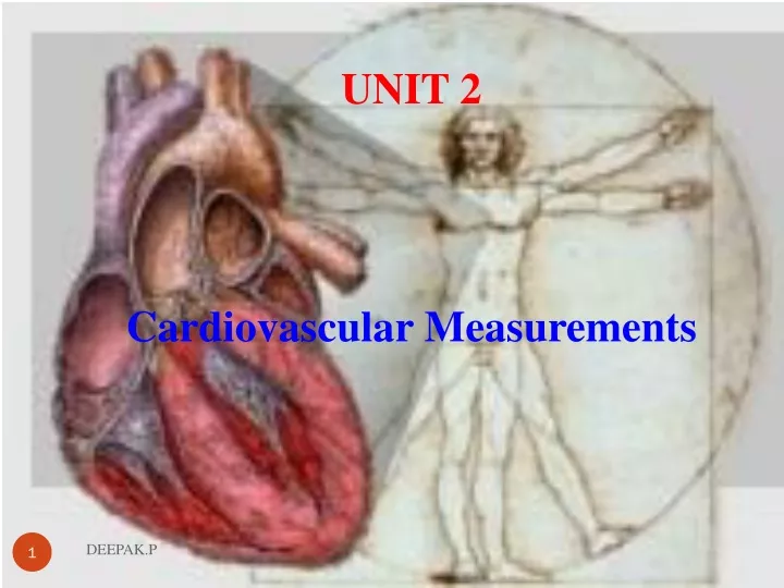
UNIT 2 Cardiovascular Measurements DEEPAK.P
Objective At the end of this Unit You will learn Different Biomedical measurements such as ECG, Blood pressure measurement, Cardiac Measurements DEEPAK.P
Cardiac Function Measurements DEEPAK.P
Measuring Cardiac Function • Blood Pressure • Electrocardiogram • Stress Test • Angiography DEEPAK.P
Measuring Cardiac Function • Blood Pressure • Measure of fluid pressure within system • Systolic Pressure: Pressure generated by contraction • Diastolic Pressure: Pressure achieved between contractions. • SBP reflects the amount of work the heart is performing • DBP indicates the amount of peripheral resistance encountered DEEPAK.P
Blood Pressure Measurement DEEPAK.P
Blood Pressure Measurements • Adequate blood pressure is essential to maintain the blood supply and function of vital organs. • A history of blood pressure measurements has saved many person from death by providing warnings of dangerously high blood pressure (hypertension) in time to provide treatment. • The maximum pressure reached during cardiac ejection is called Systole. • Minimum pressure occurring at the end of ventricular relaxation is called diastole. DEEPAK.P
Blood Pressure Measurements • In routine clinical tests, blood pressure is usually measured by means of an indirect method using a sphygmomanometer (from the Greek word, sphygmos, meaning pulse). • This method is easy to use and can be automated. • The automated indirect method of B.P measurement is called Electro sphygmomanometer DEEPAK.P
Blood Pressure Measurements • It has, however, certain disadvantages in that it does not provide a continuous recording of pressure variations and its practical repetition rate is limited. • Blood pressure is measured in millimeters of mercury (mm Hg) and recorded with the systolic number first, followed by the diastolic number. • A normal blood pressure would be recorded as 120/80 mm Hg. DEEPAK.P
Blood Pressure Measurements DEEPAK.P
Blood Pressure Measurements • The systolic pressure is the maximum pressure in an artery at the moment when the heart is beating and pumping blood through the body. • The diastolic pressure is the lowest pressure in an artery in the moments between beats when the heart is resting. • Both the systolic and diastolic pressure measurements are important • If either one is raised, it means you have high blood pressure (hypertension). DEEPAK.P
Blood Pressure Measurements • The nominal values in the basic circulatory system • Arterial system-------30-300mmHg • Venous system--------5-15mmHg • Pulmonary system----6-25mmHg • Blood pressure measurement can be classified in to • Indirect • Direct DEEPAK.P
Blood Pressure Measurements • Indirect • Simple equipment ,Very little discomfort, Less informative and Intermittent • The indirect method is also somewhat subjective, and often fails when the blood pressure is very low (as would be the case when a patient is in shock). DEEPAK.P
Indirect Blood Pressure Measurement DEEPAK.P
Blood pressure measurements • Auscultatory • Auscultatory method uses aneroid sphygmomanometer with a stethoscope. • The auscultatory method comes from the Latin word "listening. • Oscillometric • The oscillometric method was first demonstrated in 1876 and involves the observation of oscillations in the sphygmomanometer cuff pressure which are caused by the oscillations of blood flow, i.e., the pulse. DEEPAK.P
Blood pressure measurements • Palpatory • Physician identifies the flow o blood in the arteries by feeling the pulse DEEPAK.P
Blood pressure measurements using sphygmomanometer • First, a cuff is placed around your arm and inflated with a pump until the circulation is cut off. • A small valve slowly deflates the cuff, and the doctor measuring blood pressure uses a stethoscope, placed over your arm, to listen for the sound of blood pulsing through the arteries. • That first sound of rushing blood refers to the systolic blood pressure; once the sound fades, the second number indicates the diastolic pressure. DEEPAK.P
Direct Blood Pressure Measurement DEEPAK.P
Direct Blood Pressure Measurements • Provide continuous measurement • Reliable information • Transducers are directly inserted in to the blood stream • Methods for direct blood pressure measurement, on the other hand, do provide a continuous readout or recording of the blood pressure waveform and are considerably more accurate than the indirect method DEEPAK.P
Direct B.P Measurement • Methods of direct blood pressure were classified in to two • The clinical method by which the measuring device was coupled to the patient • Second, by the electrical principle involved. • First category is expanded, with the electrical principles involved being used as four subcategories. DEEPAK.P
Direct B.P Measurement • A catheterization method involving the sensing of blood pressure through a liquid column. • In this method the transducer is external to the body, and the blood pressure is transmitted through a saline solutioncolumn in a catheter to this transducer DEEPAK.P
Direct B.P Measurement • The catheterization method involving the placement of the transducer through a catheter at the actual site of measurement in the blood stream or by mounting the transducer on the tip of the catheter. • Percutaneous methods in which the blood pressure is sensed in the vessel just under the skinby the use of a needle or catheter. • Implantation techniques in which the transducer is more Permanently placed in the blood vessel or the heart by surgical methods. DEEPAK.P
B.P measurements using direct method • ln l972, Hales inserted a glass tube into the artery of a horse and crudely measured arterial pressure. • Regardless of the electrical or physical principles involved, direct measurement of blood pressure is usually obtained by one of three methods • Percutaneous insertion. • Catheterization (vessel cut down). • lmplantation of a transducer in a vessel or in the heart. DEEPAK.P
B.P measurements using direct method • ln l972, Hales inserted a glass tube into the artery of a horse and crudely measured arterial pressure. • Regardless of the electrical or physical principles involved, direct measurement of blood pressure is usually obtained by one of three methods • Percutaneous insertion. • Catheterization (vessel cut down). • Implantation of a transducer in a vessel or in the heart. DEEPAK.P
Percutaneous insertion ( direct method) • Typically, for Percutaneous insertion , a local anesthetic is injectednear the site of invasion. • The vessel is occluded and a hollow needle is inserted at a slight angle towards the vessel. • When the needle is in place, a catheter is fed through the hollow needle , usually with some sort of a guide. • When the catheter is securely place in the vessel, the needle and guide are withdrawn. DEEPAK.P
Percutaneous insertion ( direct method) • For some measurements, a type of needle attached to an airtight tube is used, so that the needle can be left in the vessel and the blood pressure sensed directly by attaching a transducer to the tube. • Other types have the transducer built in-the tip of the catheter. • This latter type is used in both percutaneous and catheterization models. DEEPAK.P
B.P measurements using direct method • ln l972, Hales inserted a glass tube into the artery of a horse and crudely measured arterial pressure. • Regardless of the electrical or physical principles involved, direct measurement of blood pressure is usually obtained by one of three methods • Percutaneous insertion. • Catheterization (vessel cut down). • lmplantation of a transducer in a vessel or in the heart. DEEPAK.P
Catheterization( direct method) • It was first developed in the late 1940s and has become a major technique for analyzing the heart and other components. • Catheter is a long tube that is inserted in to the heart or major vessels. • Sterilized catheters are used • Apart from obtaining blood pressures in the heart chamber and great vessels, this technique is also used to obtain blood samples from the heart for oxygen-content analysis and to detect the location of abnormal blood flow pathways. DEEPAK.P
Catheterization( direct method) • Measurement of blood pressure with a catheter can be achieved in two ways. • In the first method is to introduce a sterile saline solution into the catheter so that the fluid pressure is transmitted to a transducer out side the body. • In the second method, pressure measurements are obtained at the source. • Here,the transducer is introduced into the catheter and pushed to the pointat which the pressure is to be measured. or the transducer is mounted at the tip of the catheter. DEEPAK.P
Catheterization( direct method) DEEPAK.P
Catheterization( direct method) • This device is called a catheter-tip blood pressure transducer. • For mounting at the end of a catheter, one manufacturer uses an un bonded resistance strain gage in the transducer, whereas another uses a variable inductance transducer . • Implantation techniques involve major surgery. • Transducers can be categorized by the type of circuit element used to sense the pressure variations, such as capacitive, inductive, and resistive. • Since the resistive types are most frequently used. DEEPAK.P
Heart DEEPAK.P
Heart • The cardiovascular system is made of the heart, blood andblood vessels DEEPAK.P
Anatomy of the Heart • The human heart is a four-chambered muscular organ • The heart is enclosed in a pericardial bag. • The purpose of it is to protect and lubricate the heart. • The peircardium is the outermost covering of your heart. • It protects against friction rubs and protects against shocks(traumatic) as it contains 40-50 ml of pericardial fluid. • It acts as a shock absorber • . DEEPAK.P
Anatomy of the Heart • Heart normally pumps 5 liters of blood per minute • Two side of the wall is separated by the septum or dividing wall of tissue. • This septum include AV node • Right auricle is lies between inferior(lower) and superior(upper) vena cava • At the junction of Superior vena cava and right atrium SA node is situated. • . DEEPAK.P
Anatomy of the Heart • The communication between atria and ventricle is accomplished only through AV node and delay line. • The activated AV node, after a delay, initiates an impulse in to the ventricle, through the bundle of his, and bundle branches that connect to the purkinje fibers. • Ventricle wall is thicker than auricular wall • Left atrium is smaller than Right atrium • Left ventricle is considered as most important. • It wall thickness is 3 times than right ventricle. DEEPAK.P
Heart anatomy • Left heart is considered as pressure pump • Right heart is similar to a volume pump • Muscle contraction of left heart is larger and stronger than that of right heart. DEEPAK.P
Heart circulation • The work of the heart is to pump blood to the lungs through pulmonary circulation and to the rest of the body through systemic circulation. • In pulmonary circulation, the pressure difference between arteries and veins is small. • In systemic circulation, the pressure difference between arteries and veins is very high. DEEPAK.P
Heart Valves • The pumping action is accomplished by systematic contraction and relaxation of the cardiac muscle in the myocardium. • Cardiac muscles gets the blood supply from coronary circulation. • Heart contains 4 valves • Tricuspid---Between RA and RV----- Three cups • Pulmonary/Semi lunar--Between RV and Right lungs • Mitral/Bicuspid--- Between LA and LV---- Two cups • Aortic---Between LV and aorta • The sounds associated with the heartbeat are due to vibrations in the tissues and blood caused by closure of the valves. DEEPAK.P
Heart valves DEEPAK.P
Heart Sound • Listening of sound produced by heart is called auscultation • Heart sound is heard by the physician through his stethoscope. • This sound is called Korotkoff sound • The sounds associated with the heartbeat are due to vibrations in the tissues and blood caused by closure of the valves. • Normal heart produces two sounds called lub-dub • Lub is called the first heart sound • It occurs at the time of QRS complex of the ECG • Lub is related to the closure of atrioventricular valve • Which permits blood flow from auricle to ventricles. • It prevents blood flow in reverse direction DEEPAK.P
Heart Sound • Dub is called the second heart sound • Dub is related to the closure of semilunar valve • This valve releases blood into the pulmonary and systemic circulation system. • It occurs at the end of the T wave of of the ECG • Abnormal heart sounds is called murmurs. • It is due to the improper opening of the valve. • Graphic recording of heart soundis also possible • It is called phonocardiogram • Recording of the vibrations of the heart against thoracic cavity is called vibrocariogram DEEPAK.P
Cardiac Output and Rate DEEPAK.P
Cardiac Output • Cardiac output is the volume of blood pumped by the heart per minute (mL blood/min). • Cardiac output is a function of heart rate and stroke volume. • Cardiac Output in mL/min = heart rate (beats/min) X stroke volume (mL/beat) • Cardiac Output = 70 (beats/min) X 70 (mL/beat) = 4900 mL/minute. • The total volume of blood in the circulatory system of an average person is about 5 liters (5000 mL). DEEPAK.P
Cardiac Output • The heart rate is simply the number of heart beats per minute. • This can be easily measured through the use of heart rate monitors or taking ones pulse (counting the ‘pulses’ at the radial artery for example over a one minute period). • Children (ages 6 - 15) 70 – 100 beats per minute • Adults (age 18 and over) 60 – 100 beats per minute DEEPAK.P
Cardiac Output • The stroke volume is the volume of blood, in milliliters (mL), pumped out of the heart with each beat. • Strokevolume (SV) refers to the quantity of blood pumped out of the left ventricle with every heart beat. • If the volume of blood increased (waste products not being removed to the kidneys due to kidney failure for example) then there would be a greater quantity of blood within the system increasing the pressure within. DEEPAK.P
Cardiac Output • Increasing either heart rate or stroke volume increases cardiac output. DEEPAK.P